Calcium »
PDB 6sbk-6sux »
6sg8 »
Calcium in PDB 6sg8: Structure of Sosuga Virus Receptor Binding Protein
Protein crystallography data
The structure of Structure of Sosuga Virus Receptor Binding Protein, PDB code: 6sg8
was solved by
T.A.Bowden,
A.J.Stelfox,
with X-Ray Crystallography technique. A brief refinement statistics is given in the table below:
| Resolution Low / High (Å) | 51.60 / 2.50 |
| Space group | P 1 21 1 |
| Cell size a, b, c (Å), α, β, γ (°) | 75.700, 84.040, 81.690, 90.00, 112.85, 90.00 |
| R / Rfree (%) | 19.4 / 23.1 |
Calcium Binding Sites:
The binding sites of Calcium atom in the Structure of Sosuga Virus Receptor Binding Protein
(pdb code 6sg8). This binding sites where shown within
5.0 Angstroms radius around Calcium atom.
In total 2 binding sites of Calcium where determined in the Structure of Sosuga Virus Receptor Binding Protein, PDB code: 6sg8:
Jump to Calcium binding site number: 1; 2;
In total 2 binding sites of Calcium where determined in the Structure of Sosuga Virus Receptor Binding Protein, PDB code: 6sg8:
Jump to Calcium binding site number: 1; 2;
Calcium binding site 1 out of 2 in 6sg8
Go back to
Calcium binding site 1 out
of 2 in the Structure of Sosuga Virus Receptor Binding Protein
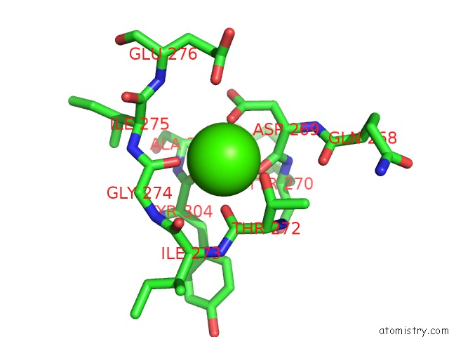
Mono view
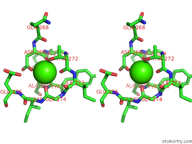
Stereo pair view
Mono view
Stereo pair view
A full contact list of Calcium with other atoms in the Ca binding
site number 1 of Structure of Sosuga Virus Receptor Binding Protein within 5.0Å range:
|
Calcium binding site 2 out of 2 in 6sg8
Go back to
Calcium binding site 2 out
of 2 in the Structure of Sosuga Virus Receptor Binding Protein
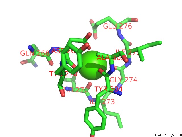
Mono view
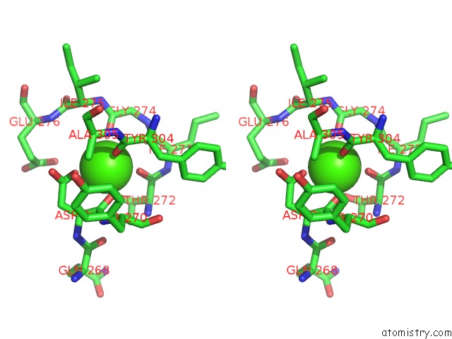
Stereo pair view
Mono view
Stereo pair view
A full contact list of Calcium with other atoms in the Ca binding
site number 2 of Structure of Sosuga Virus Receptor Binding Protein within 5.0Å range:
|
Reference:
A.J.Stelfox,
T.A.Bowden.
A Structure-Based Rationale For Sialic Acid Independent Host-Cell Entry of Sosuga Virus. Proc.Natl.Acad.Sci.Usa V. 116 21514 2019.
ISSN: ESSN 1091-6490
PubMed: 31591233
DOI: 10.1073/PNAS.1906717116
Page generated: Wed Jul 9 17:49:19 2025
ISSN: ESSN 1091-6490
PubMed: 31591233
DOI: 10.1073/PNAS.1906717116
Last articles
Mg in 6TRAMg in 6TR4
Mg in 6TR3
Mg in 6TMF
Mg in 6TQO
Mg in 6TQN
Mg in 6TQF
Mg in 6TQE
Mg in 6TQB
Mg in 6TQA