Calcium »
PDB 6sbk-6sux »
6sit »
Calcium in PDB 6sit: Pseudo-Atomic Crystal Structure of the Desmoglein 2 - Human Adenovirus Serotype 3 Fibre Knob Complex
Protein crystallography data
The structure of Pseudo-Atomic Crystal Structure of the Desmoglein 2 - Human Adenovirus Serotype 3 Fibre Knob Complex, PDB code: 6sit
was solved by
W.P.Burmeister,
P.Fender,
E.Vassal-Stermann,
with X-Ray Crystallography technique. A brief refinement statistics is given in the table below:
| Resolution Low / High (Å) | 46.50 / 4.50 |
| Space group | I 21 3 |
| Cell size a, b, c (Å), α, β, γ (°) | 146.530, 146.530, 146.530, 90.00, 90.00, 90.00 |
| R / Rfree (%) | 36.7 / 37 |
Calcium Binding Sites:
The binding sites of Calcium atom in the Pseudo-Atomic Crystal Structure of the Desmoglein 2 - Human Adenovirus Serotype 3 Fibre Knob Complex
(pdb code 6sit). This binding sites where shown within
5.0 Angstroms radius around Calcium atom.
In total 6 binding sites of Calcium where determined in the Pseudo-Atomic Crystal Structure of the Desmoglein 2 - Human Adenovirus Serotype 3 Fibre Knob Complex, PDB code: 6sit:
Jump to Calcium binding site number: 1; 2; 3; 4; 5; 6;
In total 6 binding sites of Calcium where determined in the Pseudo-Atomic Crystal Structure of the Desmoglein 2 - Human Adenovirus Serotype 3 Fibre Knob Complex, PDB code: 6sit:
Jump to Calcium binding site number: 1; 2; 3; 4; 5; 6;
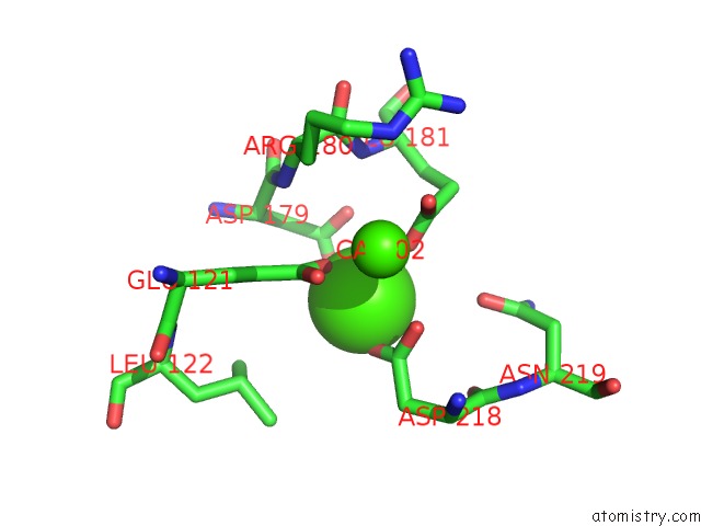
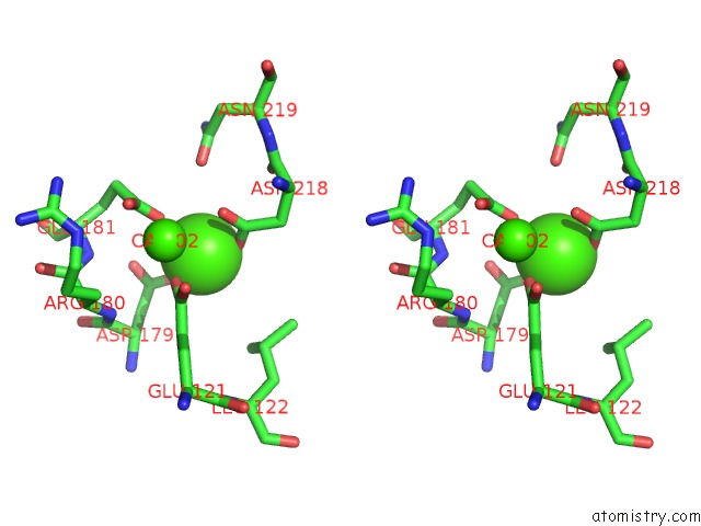
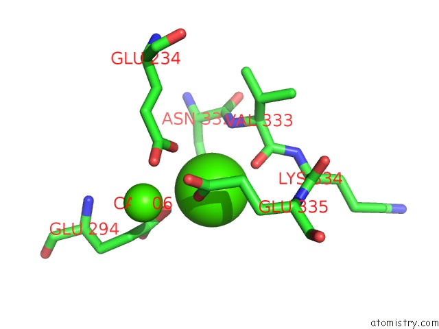
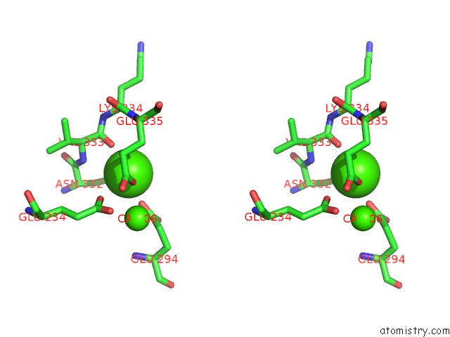
Calcium binding site 1 out of 6 in 6sit
Go back to
Calcium binding site 1 out
of 6 in the Pseudo-Atomic Crystal Structure of the Desmoglein 2 - Human Adenovirus Serotype 3 Fibre Knob Complex
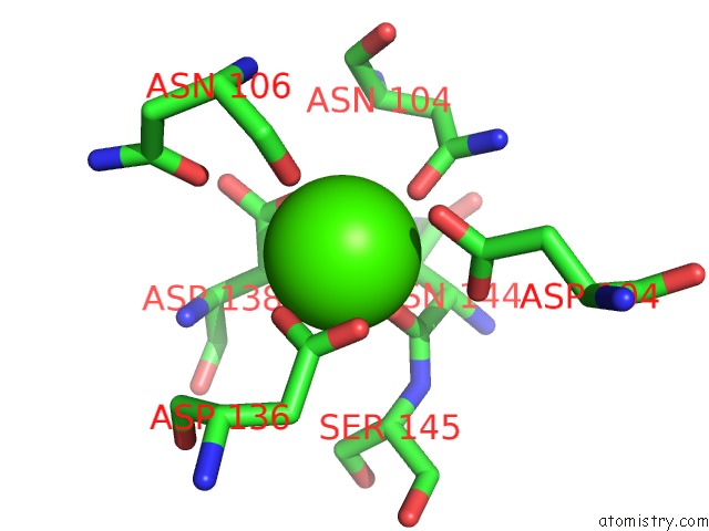
Mono view
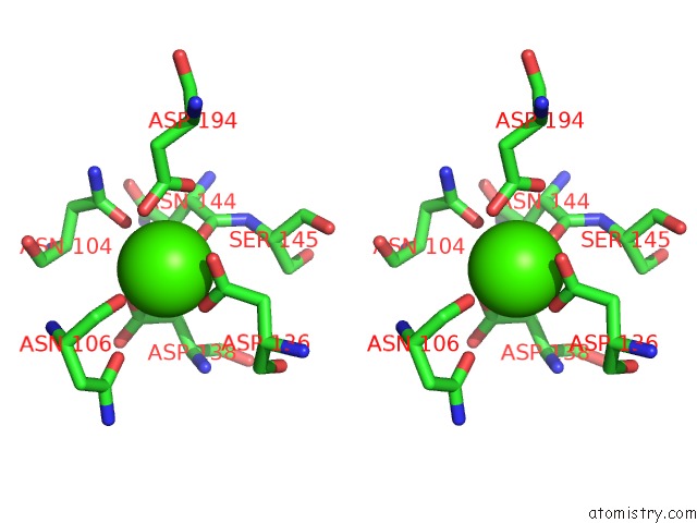
Stereo pair view
Mono view
Stereo pair view
A full contact list of Calcium with other atoms in the Ca binding
site number 1 of Pseudo-Atomic Crystal Structure of the Desmoglein 2 - Human Adenovirus Serotype 3 Fibre Knob Complex within 5.0Å range:
|
Calcium binding site 2 out of 6 in 6sit
Go back to
Calcium binding site 2 out
of 6 in the Pseudo-Atomic Crystal Structure of the Desmoglein 2 - Human Adenovirus Serotype 3 Fibre Knob Complex
Mono view
Stereo pair view
Mono view
Stereo pair view
A full contact list of Calcium with other atoms in the Ca binding
site number 2 of Pseudo-Atomic Crystal Structure of the Desmoglein 2 - Human Adenovirus Serotype 3 Fibre Knob Complex within 5.0Å range:
|
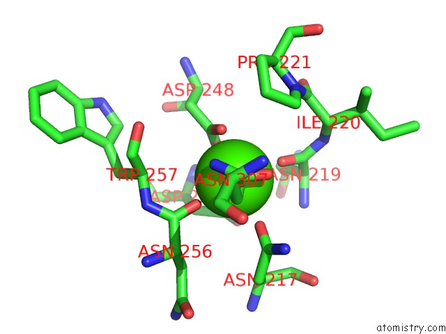
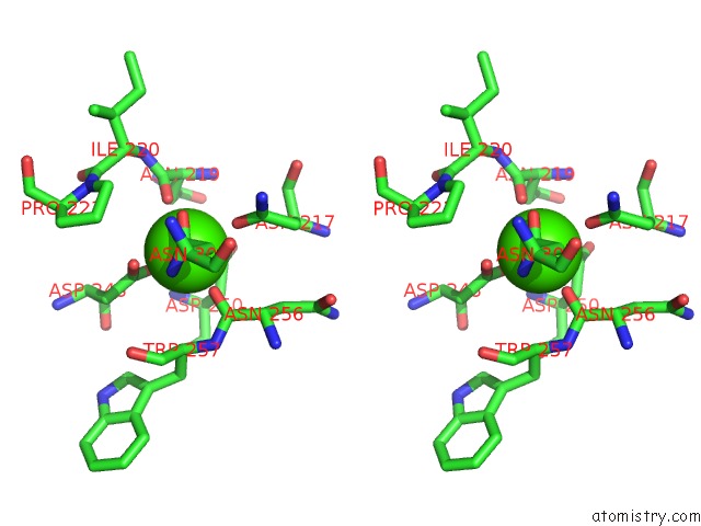
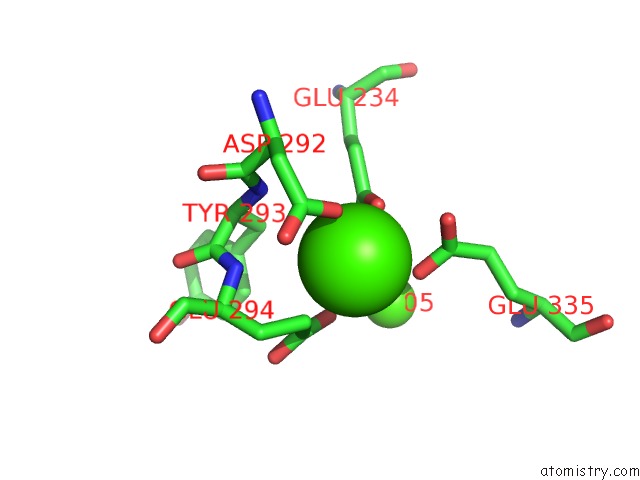
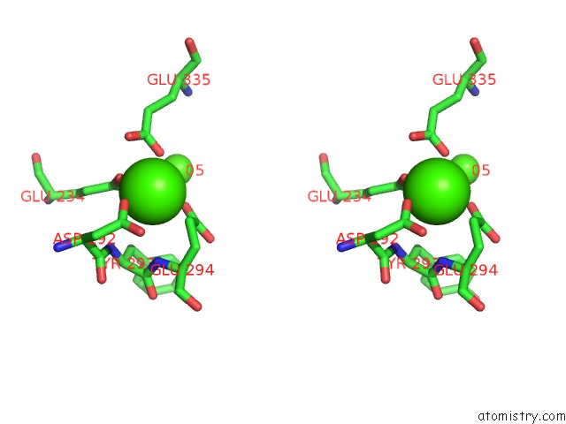
Calcium binding site 3 out of 6 in 6sit
Go back to
Calcium binding site 3 out
of 6 in the Pseudo-Atomic Crystal Structure of the Desmoglein 2 - Human Adenovirus Serotype 3 Fibre Knob Complex
Mono view
Stereo pair view
Mono view
Stereo pair view
A full contact list of Calcium with other atoms in the Ca binding
site number 3 of Pseudo-Atomic Crystal Structure of the Desmoglein 2 - Human Adenovirus Serotype 3 Fibre Knob Complex within 5.0Å range:
|
Calcium binding site 4 out of 6 in 6sit
Go back to
Calcium binding site 4 out
of 6 in the Pseudo-Atomic Crystal Structure of the Desmoglein 2 - Human Adenovirus Serotype 3 Fibre Knob Complex
Mono view
Stereo pair view
Mono view
Stereo pair view
A full contact list of Calcium with other atoms in the Ca binding
site number 4 of Pseudo-Atomic Crystal Structure of the Desmoglein 2 - Human Adenovirus Serotype 3 Fibre Knob Complex within 5.0Å range:
|
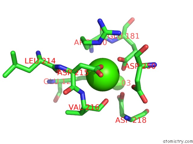
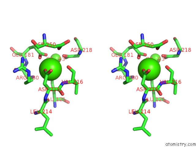
Calcium binding site 5 out of 6 in 6sit
Go back to
Calcium binding site 5 out
of 6 in the Pseudo-Atomic Crystal Structure of the Desmoglein 2 - Human Adenovirus Serotype 3 Fibre Knob Complex
Mono view
Stereo pair view
Mono view
Stereo pair view
A full contact list of Calcium with other atoms in the Ca binding
site number 5 of Pseudo-Atomic Crystal Structure of the Desmoglein 2 - Human Adenovirus Serotype 3 Fibre Knob Complex within 5.0Å range:
|
Calcium binding site 6 out of 6 in 6sit
Go back to
Calcium binding site 6 out
of 6 in the Pseudo-Atomic Crystal Structure of the Desmoglein 2 - Human Adenovirus Serotype 3 Fibre Knob Complex
Mono view
Stereo pair view
Mono view
Stereo pair view
A full contact list of Calcium with other atoms in the Ca binding
site number 6 of Pseudo-Atomic Crystal Structure of the Desmoglein 2 - Human Adenovirus Serotype 3 Fibre Knob Complex within 5.0Å range:
|
Reference:
E.Vassal-Stermann,
S.Hutin,
P.Fender,
W.P.Burmeister.
Intermediate-Resolution Crystal Structure of the Human Adenovirus B Serotype 3 Fibre Knob in Complex with the EC2-EC3 Fragment of Desmoglein 2. Acta Crystallogr.,Sect.F V. 75 750 2019.
ISSN: ESSN 2053-230X
PubMed: 31797817
DOI: 10.1107/S2053230X19015784
Page generated: Wed Jul 9 17:50:15 2025
ISSN: ESSN 2053-230X
PubMed: 31797817
DOI: 10.1107/S2053230X19015784
Last articles
Mg in 6LU1Mg in 6LT4
Mg in 6LY7
Mg in 6LY6
Mg in 6LY3
Mg in 6LX1
Mg in 6LTW
Mg in 6LVW
Mg in 6LUH
Mg in 6LTS