Calcium »
PDB 6v4s-6vgf »
6vc3 »
Calcium in PDB 6vc3: Peanut Lectin Complexed with S-Beta-D-Thiogalactopyranosyl 6-Deoxy-6- S-Propynyl-Beta-D-Glucopyranoside (Stg)
Protein crystallography data
The structure of Peanut Lectin Complexed with S-Beta-D-Thiogalactopyranosyl 6-Deoxy-6- S-Propynyl-Beta-D-Glucopyranoside (Stg), PDB code: 6vc3
was solved by
L.H.Otero,
E.D.Primo,
A.J.Cagnoni,
M.E.Cano,
S.Klinke,
F.A.Goldbaum,
M.L.Uhrig,
with X-Ray Crystallography technique. A brief refinement statistics is given in the table below:
| Resolution Low / High (Å) | 49.03 / 1.95 |
| Space group | P 2 21 21 |
| Cell size a, b, c (Å), α, β, γ (°) | 76.190, 125.418, 128.112, 90.00, 90.00, 90.00 |
| R / Rfree (%) | 26.7 / 28.3 |
Other elements in 6vc3:
The structure of Peanut Lectin Complexed with S-Beta-D-Thiogalactopyranosyl 6-Deoxy-6- S-Propynyl-Beta-D-Glucopyranoside (Stg) also contains other interesting chemical elements:
| Manganese | (Mn) | 4 atoms |
Calcium Binding Sites:
The binding sites of Calcium atom in the Peanut Lectin Complexed with S-Beta-D-Thiogalactopyranosyl 6-Deoxy-6- S-Propynyl-Beta-D-Glucopyranoside (Stg)
(pdb code 6vc3). This binding sites where shown within
5.0 Angstroms radius around Calcium atom.
In total 4 binding sites of Calcium where determined in the Peanut Lectin Complexed with S-Beta-D-Thiogalactopyranosyl 6-Deoxy-6- S-Propynyl-Beta-D-Glucopyranoside (Stg), PDB code: 6vc3:
Jump to Calcium binding site number: 1; 2; 3; 4;
In total 4 binding sites of Calcium where determined in the Peanut Lectin Complexed with S-Beta-D-Thiogalactopyranosyl 6-Deoxy-6- S-Propynyl-Beta-D-Glucopyranoside (Stg), PDB code: 6vc3:
Jump to Calcium binding site number: 1; 2; 3; 4;
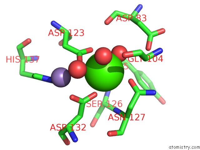
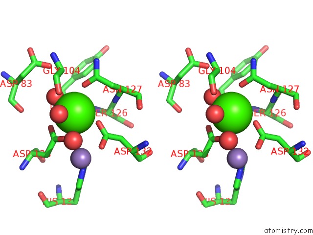
Calcium binding site 1 out of 4 in 6vc3
Go back to
Calcium binding site 1 out
of 4 in the Peanut Lectin Complexed with S-Beta-D-Thiogalactopyranosyl 6-Deoxy-6- S-Propynyl-Beta-D-Glucopyranoside (Stg)
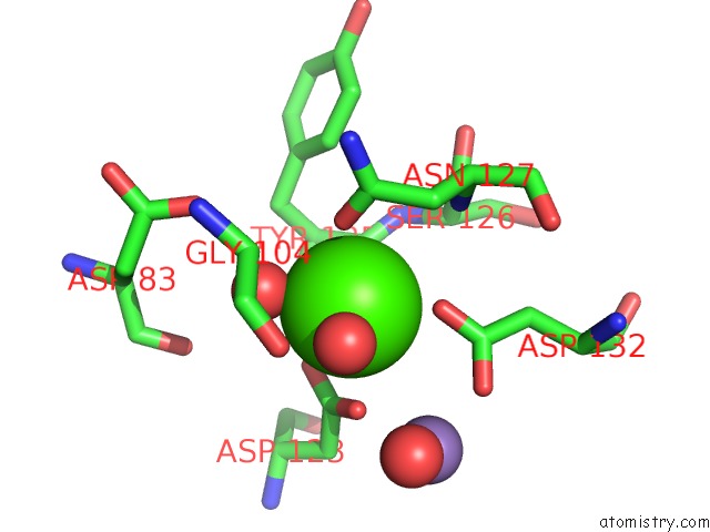
Mono view
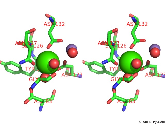
Stereo pair view
Mono view
Stereo pair view
A full contact list of Calcium with other atoms in the Ca binding
site number 1 of Peanut Lectin Complexed with S-Beta-D-Thiogalactopyranosyl 6-Deoxy-6- S-Propynyl-Beta-D-Glucopyranoside (Stg) within 5.0Å range:
|
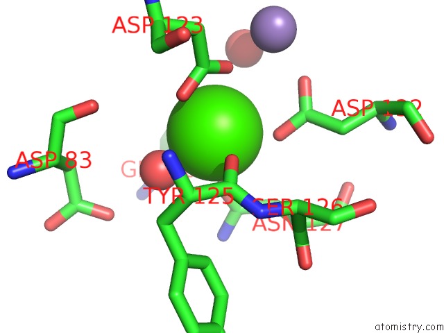
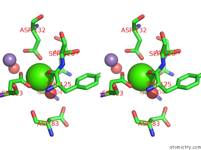
Calcium binding site 2 out of 4 in 6vc3
Go back to
Calcium binding site 2 out
of 4 in the Peanut Lectin Complexed with S-Beta-D-Thiogalactopyranosyl 6-Deoxy-6- S-Propynyl-Beta-D-Glucopyranoside (Stg)
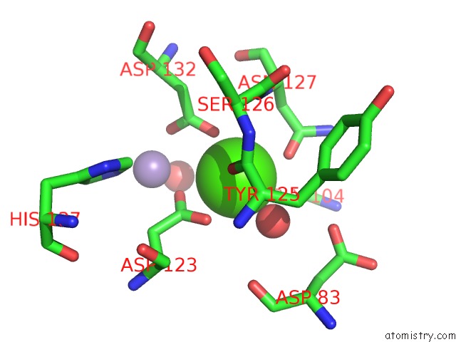
Mono view
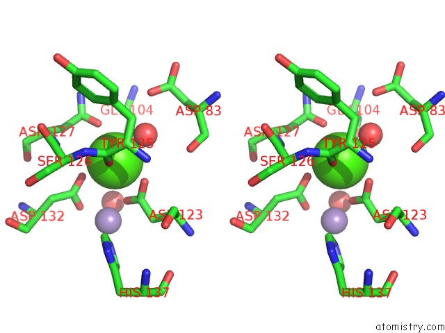
Stereo pair view
Mono view
Stereo pair view
A full contact list of Calcium with other atoms in the Ca binding
site number 2 of Peanut Lectin Complexed with S-Beta-D-Thiogalactopyranosyl 6-Deoxy-6- S-Propynyl-Beta-D-Glucopyranoside (Stg) within 5.0Å range:
|
Calcium binding site 3 out of 4 in 6vc3
Go back to
Calcium binding site 3 out
of 4 in the Peanut Lectin Complexed with S-Beta-D-Thiogalactopyranosyl 6-Deoxy-6- S-Propynyl-Beta-D-Glucopyranoside (Stg)
Mono view
Stereo pair view
Mono view
Stereo pair view
A full contact list of Calcium with other atoms in the Ca binding
site number 3 of Peanut Lectin Complexed with S-Beta-D-Thiogalactopyranosyl 6-Deoxy-6- S-Propynyl-Beta-D-Glucopyranoside (Stg) within 5.0Å range:
|
Calcium binding site 4 out of 4 in 6vc3
Go back to
Calcium binding site 4 out
of 4 in the Peanut Lectin Complexed with S-Beta-D-Thiogalactopyranosyl 6-Deoxy-6- S-Propynyl-Beta-D-Glucopyranoside (Stg)
Mono view
Stereo pair view
Mono view
Stereo pair view
A full contact list of Calcium with other atoms in the Ca binding
site number 4 of Peanut Lectin Complexed with S-Beta-D-Thiogalactopyranosyl 6-Deoxy-6- S-Propynyl-Beta-D-Glucopyranoside (Stg) within 5.0Å range:
|
Reference:
A.J.Cagnoni,
E.D.Primo,
S.Klinke,
M.E.Cano,
W.Giordano,
K.V.Marino,
J.Kovensky,
F.A.Goldbaum,
M.L.Uhrig,
L.H.Otero.
Crystal Structures of Peanut Lectin in the Presence of Synthetic Beta-N- and Beta-S-Galactosides Disclose Evidence For the Recognition of Different Glycomimetic Ligands. Acta Crystallogr D Struct V. 76 1080 2020BIOL.
ISSN: ISSN 2059-7983
PubMed: 33135679
DOI: 10.1107/S2059798320012371
Page generated: Wed Jul 9 19:03:36 2025
ISSN: ISSN 2059-7983
PubMed: 33135679
DOI: 10.1107/S2059798320012371
Last articles
K in 7QR0K in 7QR1
K in 7QQZ
K in 7QQX
K in 7QQW
K in 7QQU
K in 7QQV
K in 7QQT
K in 7QQR
K in 7QQS