Calcium »
PDB 6v4s-6vgf »
6vel »
Calcium in PDB 6vel: Crystal Structure of Human E-Cadherin Bound By Mouse Monoclonal Antibody 66E8FAB
Protein crystallography data
The structure of Crystal Structure of Human E-Cadherin Bound By Mouse Monoclonal Antibody 66E8FAB, PDB code: 6vel
was solved by
Seattle Structural Genomics Center For Infectious Disease,
Seattlestructural Genomics Center For Infectious Disease (Ssgcid),
with X-Ray Crystallography technique. A brief refinement statistics is given in the table below:
| Resolution Low / High (Å) | 46.54 / 2.65 |
| Space group | P 31 2 1 |
| Cell size a, b, c (Å), α, β, γ (°) | 142.170, 142.170, 90.320, 90.00, 90.00, 120.00 |
| R / Rfree (%) | 18.3 / 23.3 |
Other elements in 6vel:
The structure of Crystal Structure of Human E-Cadherin Bound By Mouse Monoclonal Antibody 66E8FAB also contains other interesting chemical elements:
| Sodium | (Na) | 1 atom |
Calcium Binding Sites:
The binding sites of Calcium atom in the Crystal Structure of Human E-Cadherin Bound By Mouse Monoclonal Antibody 66E8FAB
(pdb code 6vel). This binding sites where shown within
5.0 Angstroms radius around Calcium atom.
In total 2 binding sites of Calcium where determined in the Crystal Structure of Human E-Cadherin Bound By Mouse Monoclonal Antibody 66E8FAB, PDB code: 6vel:
Jump to Calcium binding site number: 1; 2;
In total 2 binding sites of Calcium where determined in the Crystal Structure of Human E-Cadherin Bound By Mouse Monoclonal Antibody 66E8FAB, PDB code: 6vel:
Jump to Calcium binding site number: 1; 2;
Calcium binding site 1 out of 2 in 6vel
Go back to
Calcium binding site 1 out
of 2 in the Crystal Structure of Human E-Cadherin Bound By Mouse Monoclonal Antibody 66E8FAB

Mono view
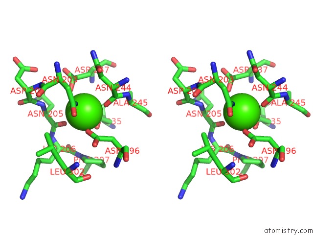
Stereo pair view
Mono view
Stereo pair view
A full contact list of Calcium with other atoms in the Ca binding
site number 1 of Crystal Structure of Human E-Cadherin Bound By Mouse Monoclonal Antibody 66E8FAB within 5.0Å range:
|
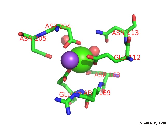
Calcium binding site 2 out of 2 in 6vel
Go back to
Calcium binding site 2 out
of 2 in the Crystal Structure of Human E-Cadherin Bound By Mouse Monoclonal Antibody 66E8FAB
Mono view
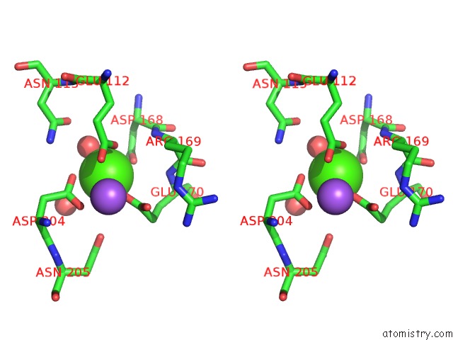
Stereo pair view
Mono view
Stereo pair view
A full contact list of Calcium with other atoms in the Ca binding
site number 2 of Crystal Structure of Human E-Cadherin Bound By Mouse Monoclonal Antibody 66E8FAB within 5.0Å range:
|
Reference:
M.J.Bolejack,
J.Abendroth,
A.Y.Maker,
B.M.Gumbiner,
D.D.Lorimer,
P.S.Horanyi,
T.E.Edwards.
Crystal Structure of Human E-Cadherin Bound By Mouse Monoclonal Antibody 66E8FAB To Be Published.
Page generated: Wed Jul 9 19:03:38 2025
Last articles
K in 7M2HK in 7MDJ
K in 7MJQ
K in 7MHR
K in 7MJO
K in 7M0D
K in 7M89
K in 7M83
K in 7M2I
K in 7M7T