Calcium »
PDB 6wni-6x6q »
6x0i »
Calcium in PDB 6x0i: Structure of Oxidized Sida Ornithine Hydroxylase with the Fad "in" and Complexed with Nadp
Enzymatic activity of Structure of Oxidized Sida Ornithine Hydroxylase with the Fad "in" and Complexed with Nadp
All present enzymatic activity of Structure of Oxidized Sida Ornithine Hydroxylase with the Fad "in" and Complexed with Nadp:
1.14.13.196;
1.14.13.196;
Protein crystallography data
The structure of Structure of Oxidized Sida Ornithine Hydroxylase with the Fad "in" and Complexed with Nadp, PDB code: 6x0i
was solved by
J.J.Tanner,
A.C.Campbell,
with X-Ray Crystallography technique. A brief refinement statistics is given in the table below:
| Resolution Low / High (Å) | 63.21 / 1.95 |
| Space group | P 1 21 1 |
| Cell size a, b, c (Å), α, β, γ (°) | 80.507, 154.868, 90.483, 90.00, 109.25, 90.00 |
| R / Rfree (%) | 16.8 / 21.1 |
Calcium Binding Sites:
The binding sites of Calcium atom in the Structure of Oxidized Sida Ornithine Hydroxylase with the Fad "in" and Complexed with Nadp
(pdb code 6x0i). This binding sites where shown within
5.0 Angstroms radius around Calcium atom.
In total 4 binding sites of Calcium where determined in the Structure of Oxidized Sida Ornithine Hydroxylase with the Fad "in" and Complexed with Nadp, PDB code: 6x0i:
Jump to Calcium binding site number: 1; 2; 3; 4;
In total 4 binding sites of Calcium where determined in the Structure of Oxidized Sida Ornithine Hydroxylase with the Fad "in" and Complexed with Nadp, PDB code: 6x0i:
Jump to Calcium binding site number: 1; 2; 3; 4;
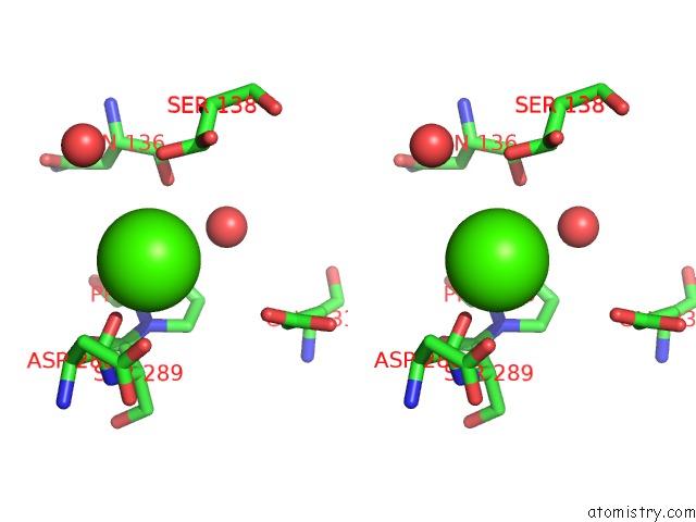
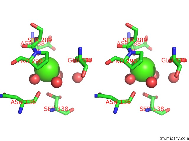
Calcium binding site 1 out of 4 in 6x0i
Go back to
Calcium binding site 1 out
of 4 in the Structure of Oxidized Sida Ornithine Hydroxylase with the Fad "in" and Complexed with Nadp

Mono view
Stereo pair view
Mono view
Stereo pair view
A full contact list of Calcium with other atoms in the Ca binding
site number 1 of Structure of Oxidized Sida Ornithine Hydroxylase with the Fad "in" and Complexed with Nadp within 5.0Å range:
|
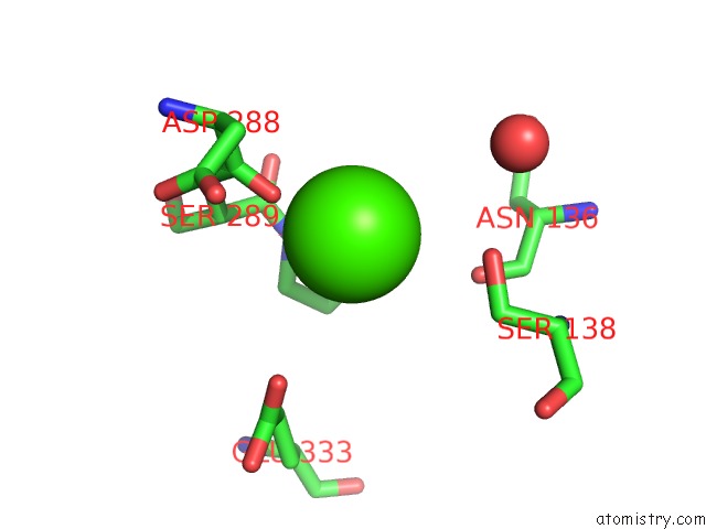
Calcium binding site 2 out of 4 in 6x0i
Go back to
Calcium binding site 2 out
of 4 in the Structure of Oxidized Sida Ornithine Hydroxylase with the Fad "in" and Complexed with Nadp

Mono view

Stereo pair view
Mono view
Stereo pair view
A full contact list of Calcium with other atoms in the Ca binding
site number 2 of Structure of Oxidized Sida Ornithine Hydroxylase with the Fad "in" and Complexed with Nadp within 5.0Å range:
|
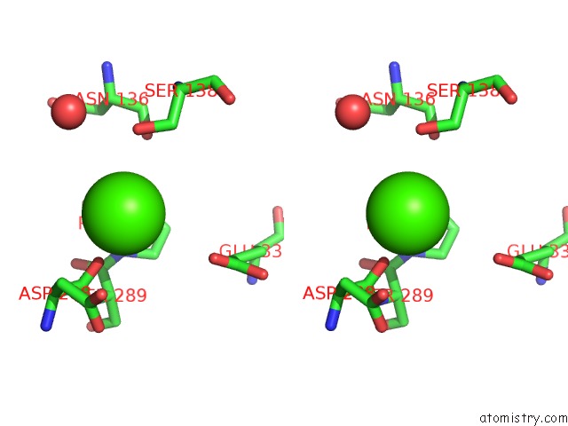
Calcium binding site 3 out of 4 in 6x0i
Go back to
Calcium binding site 3 out
of 4 in the Structure of Oxidized Sida Ornithine Hydroxylase with the Fad "in" and Complexed with Nadp
Mono view
Stereo pair view
Mono view
Stereo pair view
A full contact list of Calcium with other atoms in the Ca binding
site number 3 of Structure of Oxidized Sida Ornithine Hydroxylase with the Fad "in" and Complexed with Nadp within 5.0Å range:
|
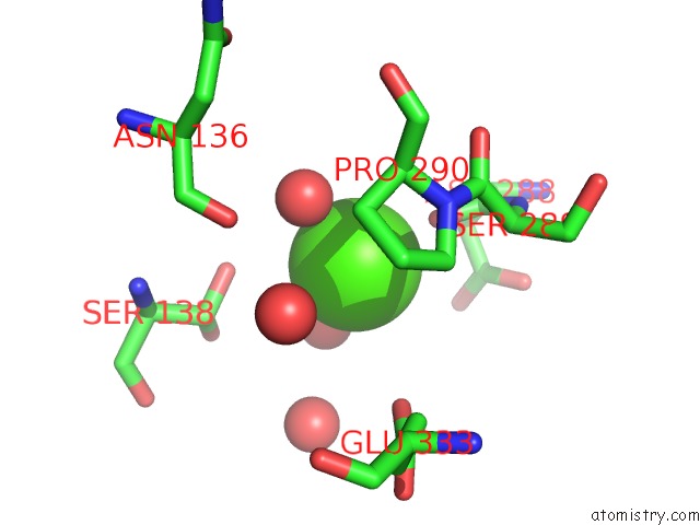
Calcium binding site 4 out of 4 in 6x0i
Go back to
Calcium binding site 4 out
of 4 in the Structure of Oxidized Sida Ornithine Hydroxylase with the Fad "in" and Complexed with Nadp
Mono view
Stereo pair view
Mono view
Stereo pair view
A full contact list of Calcium with other atoms in the Ca binding
site number 4 of Structure of Oxidized Sida Ornithine Hydroxylase with the Fad "in" and Complexed with Nadp within 5.0Å range:
|
Reference:
A.C.Campbell,
K.M.Stiers,
J.S.Martin Del Campo,
R.Mehra-Chaudhary,
P.Sobrado,
J.J.Tanner.
Trapping Conformational States of A Flavin-Dependent N -Monooxygenase in Crystallo Reveals Protein and Flavin Dynamics. J.Biol.Chem. V. 295 13239 2020.
ISSN: ESSN 1083-351X
PubMed: 32723870
DOI: 10.1074/JBC.RA120.014750
Page generated: Wed Jul 9 19:44:31 2025
ISSN: ESSN 1083-351X
PubMed: 32723870
DOI: 10.1074/JBC.RA120.014750
Last articles
Mg in 6YBWMg in 6YKW
Mg in 6YKV
Mg in 6YKU
Mg in 6YKT
Mg in 6YKS
Mg in 6YKQ
Mg in 6YKO
Mg in 6YKN
Mg in 6YKL