Calcium »
PDB 6x6z-6xrv »
6x8y »
Calcium in PDB 6x8y: Ribose-Bound Structure of Marinomonas Primoryensis PA14 Carbohydrate- Binding Domain
Protein crystallography data
The structure of Ribose-Bound Structure of Marinomonas Primoryensis PA14 Carbohydrate- Binding Domain, PDB code: 6x8y
was solved by
S.Guo,
P.L.Davies,
with X-Ray Crystallography technique. A brief refinement statistics is given in the table below:
| Resolution Low / High (Å) | 42.78 / 1.00 |
| Space group | P 21 21 21 |
| Cell size a, b, c (Å), α, β, γ (°) | 45.14, 50.72, 79.61, 90, 90, 90 |
| R / Rfree (%) | 15.5 / 16.7 |
Calcium Binding Sites:
The binding sites of Calcium atom in the Ribose-Bound Structure of Marinomonas Primoryensis PA14 Carbohydrate- Binding Domain
(pdb code 6x8y). This binding sites where shown within
5.0 Angstroms radius around Calcium atom.
In total 4 binding sites of Calcium where determined in the Ribose-Bound Structure of Marinomonas Primoryensis PA14 Carbohydrate- Binding Domain, PDB code: 6x8y:
Jump to Calcium binding site number: 1; 2; 3; 4;
In total 4 binding sites of Calcium where determined in the Ribose-Bound Structure of Marinomonas Primoryensis PA14 Carbohydrate- Binding Domain, PDB code: 6x8y:
Jump to Calcium binding site number: 1; 2; 3; 4;
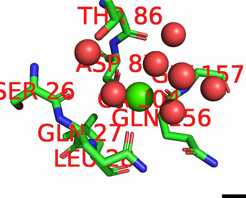
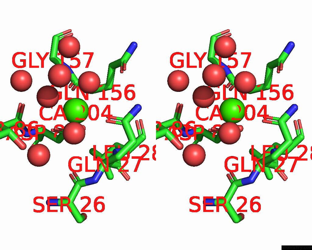
Calcium binding site 1 out of 4 in 6x8y
Go back to
Calcium binding site 1 out
of 4 in the Ribose-Bound Structure of Marinomonas Primoryensis PA14 Carbohydrate- Binding Domain
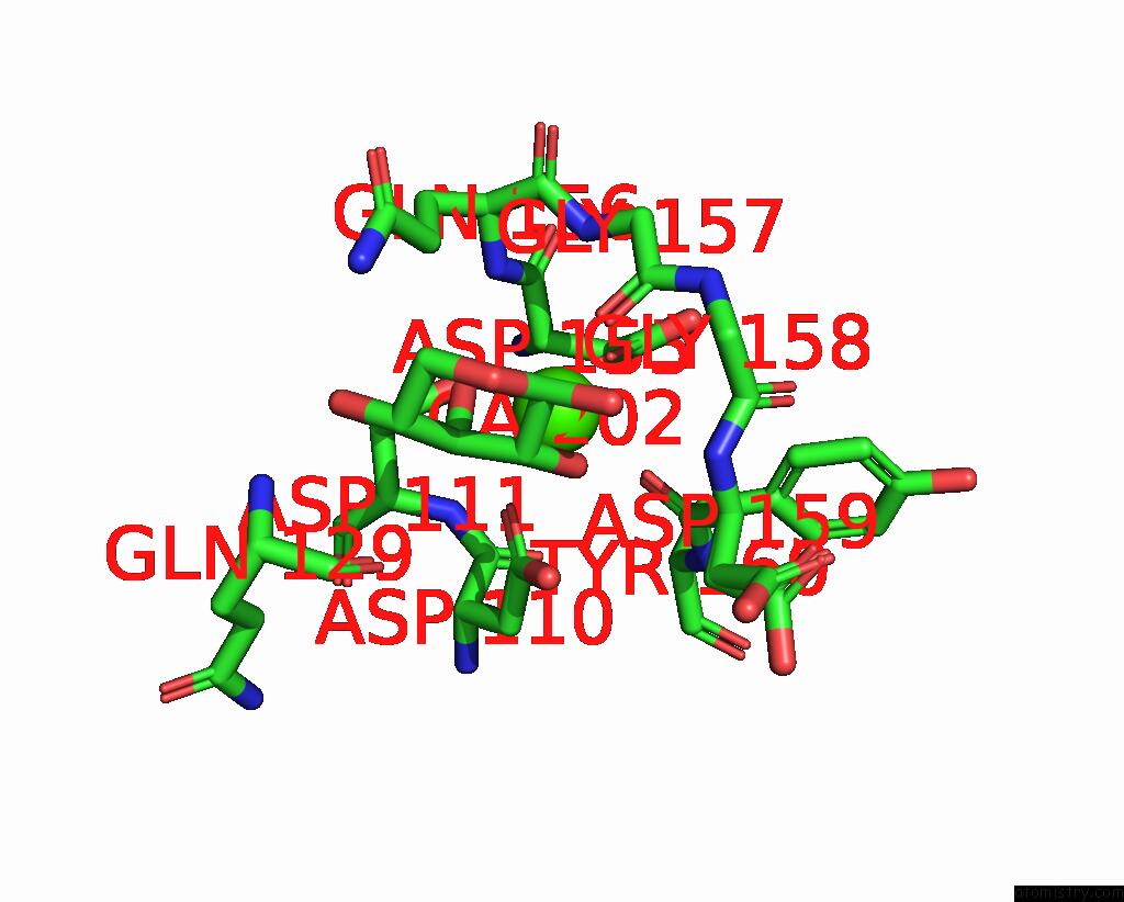
Mono view
Stereo pair view
Mono view
Stereo pair view
A full contact list of Calcium with other atoms in the Ca binding
site number 1 of Ribose-Bound Structure of Marinomonas Primoryensis PA14 Carbohydrate- Binding Domain within 5.0Å range:
|
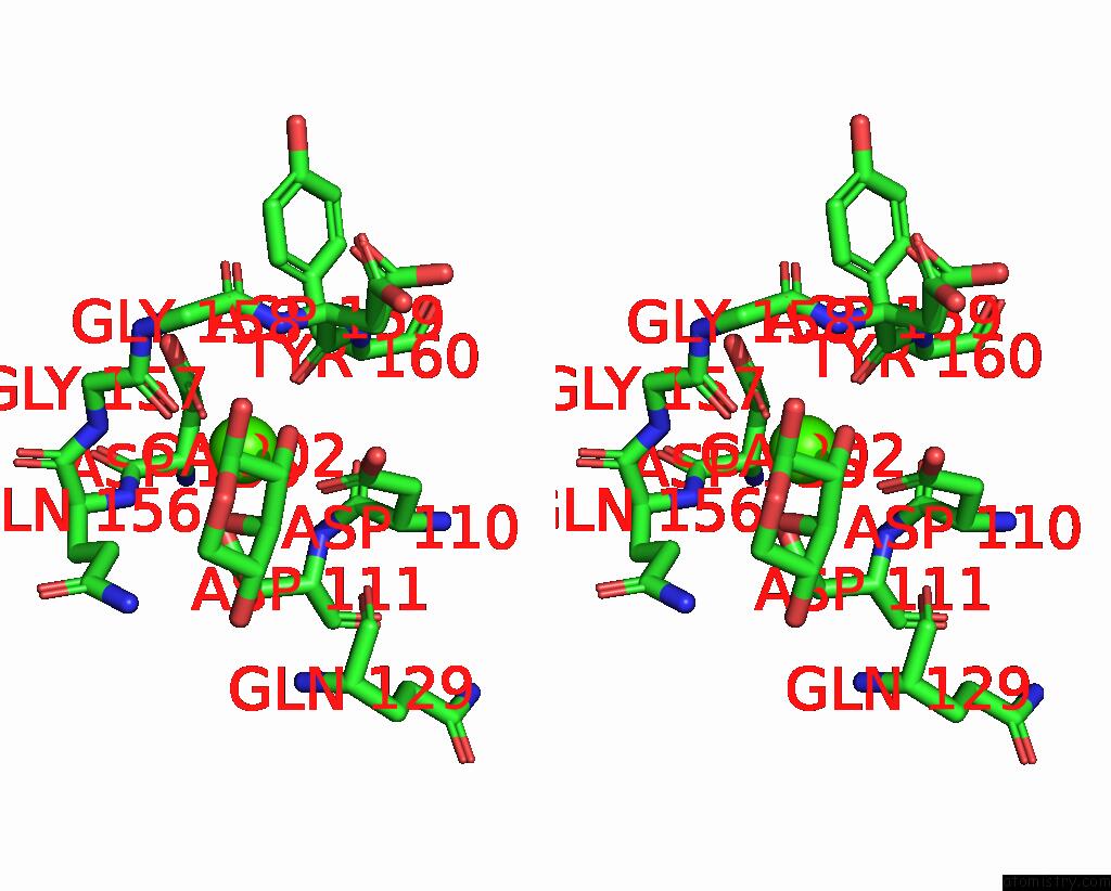
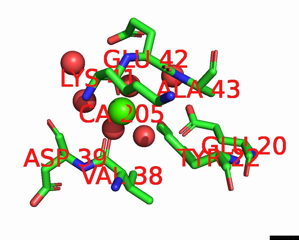
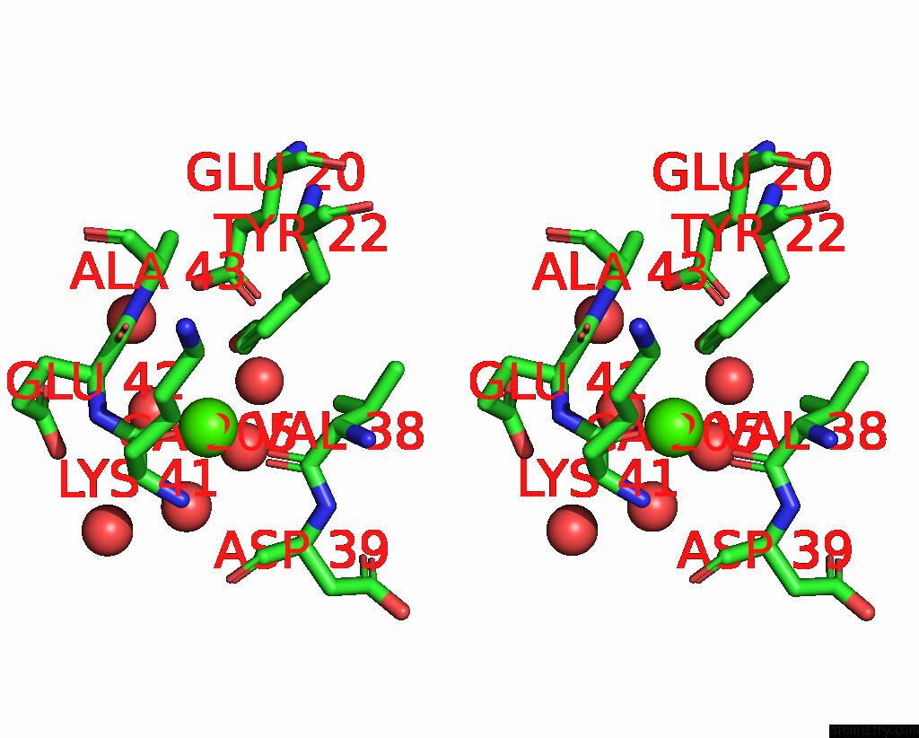
Calcium binding site 2 out of 4 in 6x8y
Go back to
Calcium binding site 2 out
of 4 in the Ribose-Bound Structure of Marinomonas Primoryensis PA14 Carbohydrate- Binding Domain
Mono view
Stereo pair view
Mono view
Stereo pair view
A full contact list of Calcium with other atoms in the Ca binding
site number 2 of Ribose-Bound Structure of Marinomonas Primoryensis PA14 Carbohydrate- Binding Domain within 5.0Å range:
|
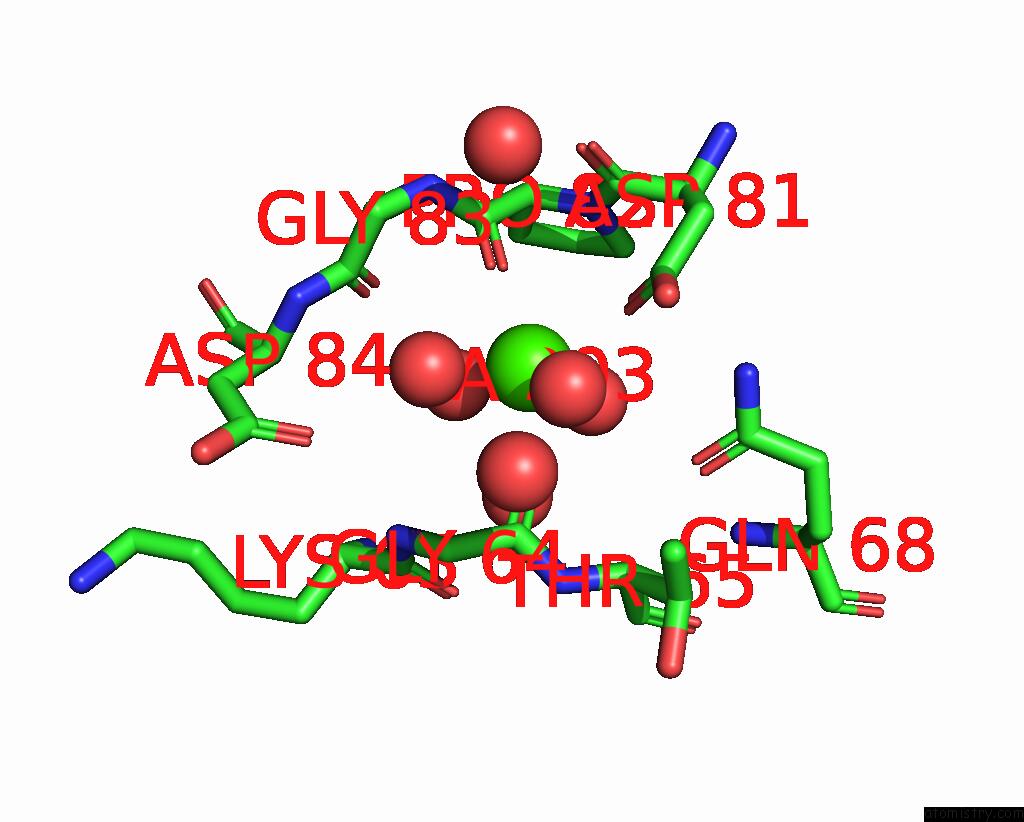
Calcium binding site 3 out of 4 in 6x8y
Go back to
Calcium binding site 3 out
of 4 in the Ribose-Bound Structure of Marinomonas Primoryensis PA14 Carbohydrate- Binding Domain
Mono view
Stereo pair view
Mono view
Stereo pair view
A full contact list of Calcium with other atoms in the Ca binding
site number 3 of Ribose-Bound Structure of Marinomonas Primoryensis PA14 Carbohydrate- Binding Domain within 5.0Å range:
|
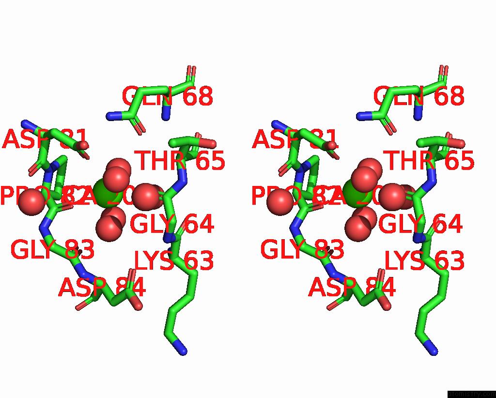
Calcium binding site 4 out of 4 in 6x8y
Go back to
Calcium binding site 4 out
of 4 in the Ribose-Bound Structure of Marinomonas Primoryensis PA14 Carbohydrate- Binding Domain
Mono view
Stereo pair view
Mono view
Stereo pair view
A full contact list of Calcium with other atoms in the Ca binding
site number 4 of Ribose-Bound Structure of Marinomonas Primoryensis PA14 Carbohydrate- Binding Domain within 5.0Å range:
|
Reference:
S.Guo,
T.D.R.Vance,
H.Zahiri,
R.Eves,
C.Stevens,
J.H.Hehemann,
S.Vidal-Melgosa,
P.L.Davies.
Structural Basis of Ligand Selectivity By A Bacterial Adhesin Lectin Involved in Multispecies Biofilm Formation. Mbio V. 12 2021.
ISSN: ESSN 2150-7511
PubMed: 33824212
DOI: 10.1128/MBIO.00130-21
Page generated: Wed Jul 9 19:55:49 2025
ISSN: ESSN 2150-7511
PubMed: 33824212
DOI: 10.1128/MBIO.00130-21
Last articles
Ir in 5T83Ir in 5K7D
Ir in 5E1U
Ir in 5JMG
Ir in 5HQO
Ir in 5E2D
Ir in 4R0D
Ir in 4Y1N
Ir in 5DE8
Ir in 5BRV