Calcium »
PDB 6xs3-6y43 »
6xw2 »
Calcium in PDB 6xw2: Crystal Structure of the Bright Genetically Encoded Calcium Indicator NCAMP7 Based on Mneongreen Fluorescent Protein
Protein crystallography data
The structure of Crystal Structure of the Bright Genetically Encoded Calcium Indicator NCAMP7 Based on Mneongreen Fluorescent Protein, PDB code: 6xw2
was solved by
K.M.Boyko,
A.Y.Nikolaeva,
D.A.Korzhenevskiy,
V.A.Lazarenko,
O.M.Subach,
F.V.Subach,
with X-Ray Crystallography technique. A brief refinement statistics is given in the table below:
| Resolution Low / High (Å) | 37.99 / 1.75 |
| Space group | P 21 21 21 |
| Cell size a, b, c (Å), α, β, γ (°) | 61.900, 65.700, 93.020, 90.00, 90.00, 90.00 |
| R / Rfree (%) | 16.3 / 19.3 |
Calcium Binding Sites:
The binding sites of Calcium atom in the Crystal Structure of the Bright Genetically Encoded Calcium Indicator NCAMP7 Based on Mneongreen Fluorescent Protein
(pdb code 6xw2). This binding sites where shown within
5.0 Angstroms radius around Calcium atom.
In total 4 binding sites of Calcium where determined in the Crystal Structure of the Bright Genetically Encoded Calcium Indicator NCAMP7 Based on Mneongreen Fluorescent Protein, PDB code: 6xw2:
Jump to Calcium binding site number: 1; 2; 3; 4;
In total 4 binding sites of Calcium where determined in the Crystal Structure of the Bright Genetically Encoded Calcium Indicator NCAMP7 Based on Mneongreen Fluorescent Protein, PDB code: 6xw2:
Jump to Calcium binding site number: 1; 2; 3; 4;
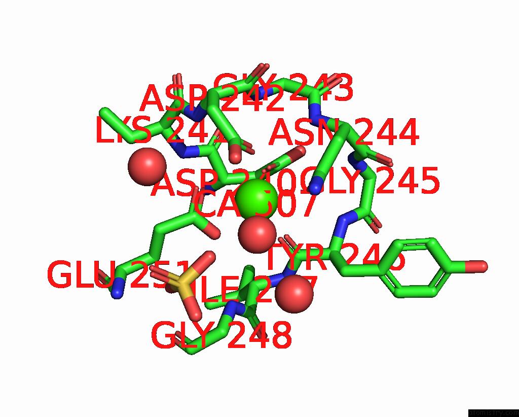
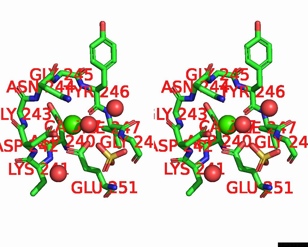
Calcium binding site 1 out of 4 in 6xw2
Go back to
Calcium binding site 1 out
of 4 in the Crystal Structure of the Bright Genetically Encoded Calcium Indicator NCAMP7 Based on Mneongreen Fluorescent Protein
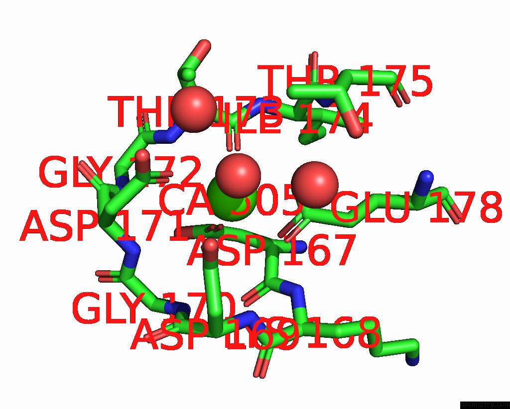
Mono view
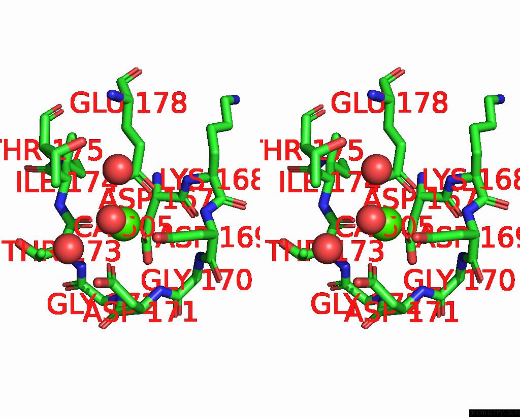
Stereo pair view
Mono view
Stereo pair view
A full contact list of Calcium with other atoms in the Ca binding
site number 1 of Crystal Structure of the Bright Genetically Encoded Calcium Indicator NCAMP7 Based on Mneongreen Fluorescent Protein within 5.0Å range:
|
Calcium binding site 2 out of 4 in 6xw2
Go back to
Calcium binding site 2 out
of 4 in the Crystal Structure of the Bright Genetically Encoded Calcium Indicator NCAMP7 Based on Mneongreen Fluorescent Protein
Mono view
Stereo pair view
Mono view
Stereo pair view
A full contact list of Calcium with other atoms in the Ca binding
site number 2 of Crystal Structure of the Bright Genetically Encoded Calcium Indicator NCAMP7 Based on Mneongreen Fluorescent Protein within 5.0Å range:
|
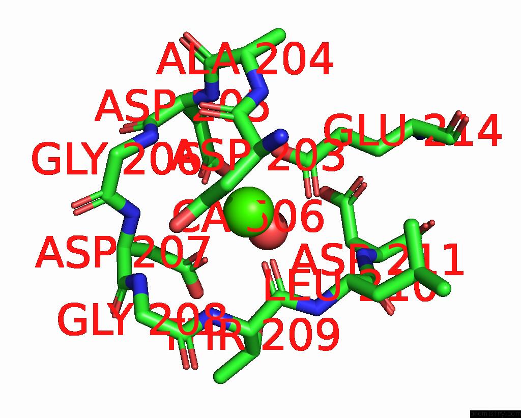
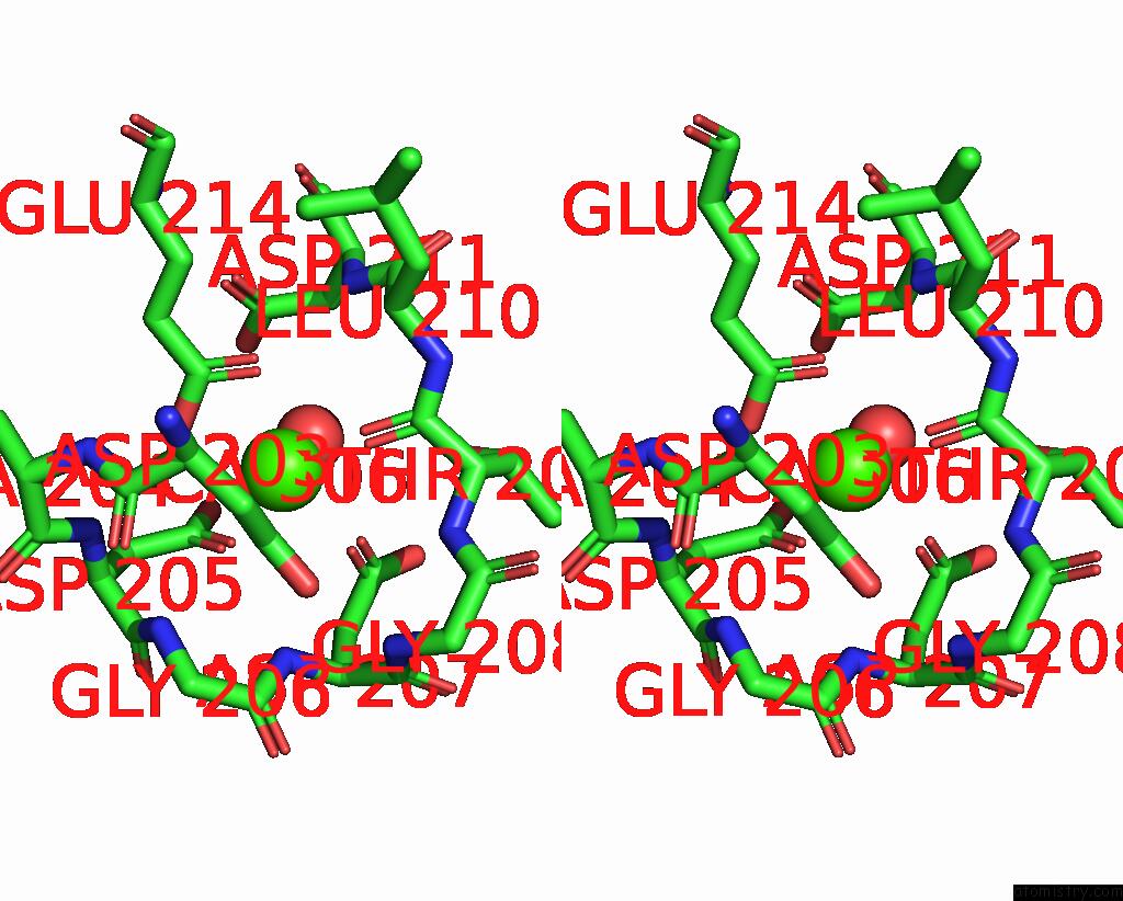
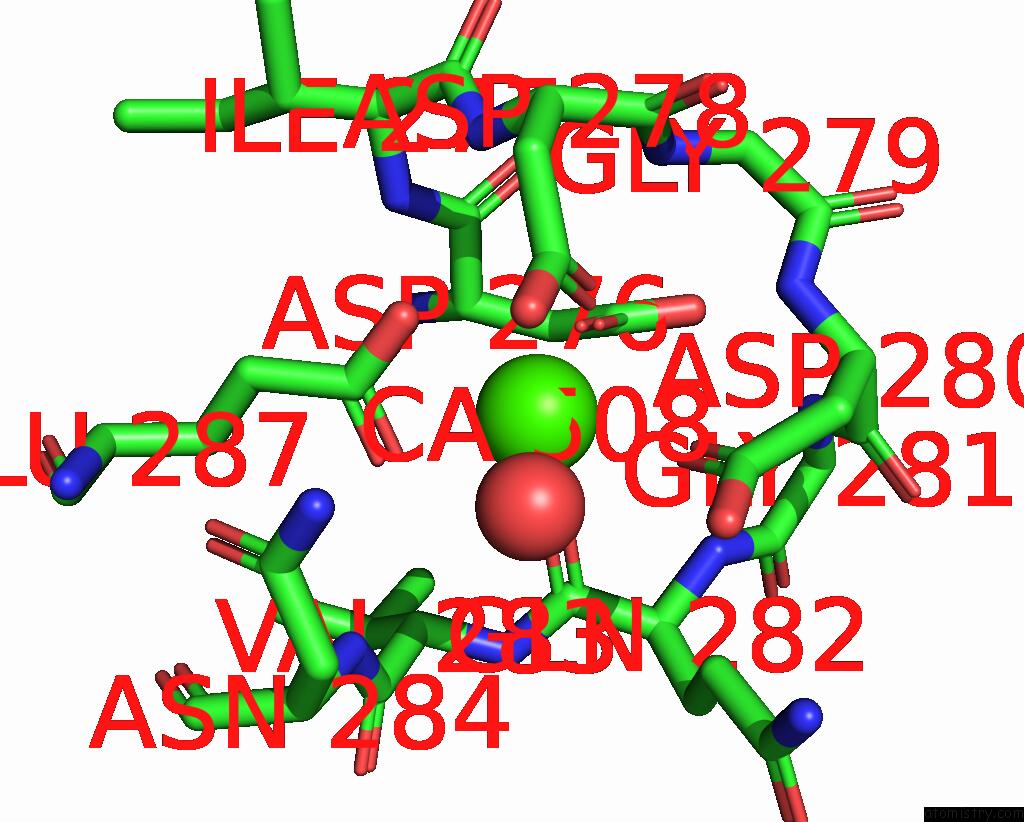
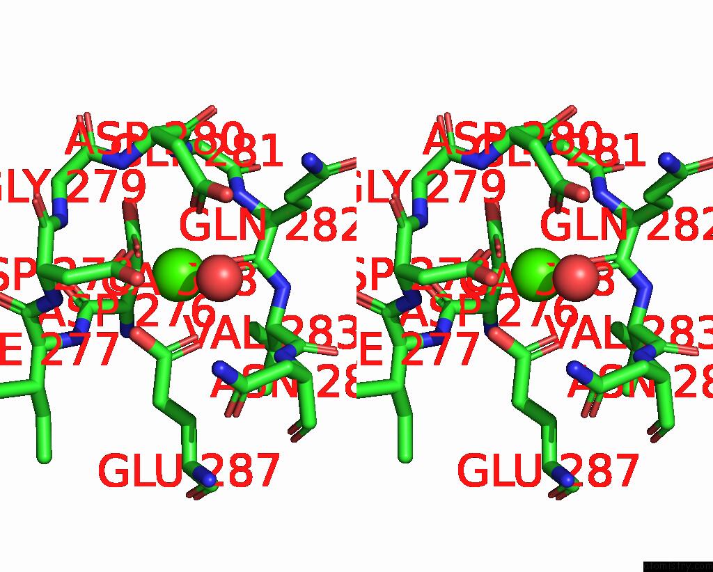
Calcium binding site 3 out of 4 in 6xw2
Go back to
Calcium binding site 3 out
of 4 in the Crystal Structure of the Bright Genetically Encoded Calcium Indicator NCAMP7 Based on Mneongreen Fluorescent Protein
Mono view
Stereo pair view
Mono view
Stereo pair view
A full contact list of Calcium with other atoms in the Ca binding
site number 3 of Crystal Structure of the Bright Genetically Encoded Calcium Indicator NCAMP7 Based on Mneongreen Fluorescent Protein within 5.0Å range:
|
Calcium binding site 4 out of 4 in 6xw2
Go back to
Calcium binding site 4 out
of 4 in the Crystal Structure of the Bright Genetically Encoded Calcium Indicator NCAMP7 Based on Mneongreen Fluorescent Protein
Mono view
Stereo pair view
Mono view
Stereo pair view
A full contact list of Calcium with other atoms in the Ca binding
site number 4 of Crystal Structure of the Bright Genetically Encoded Calcium Indicator NCAMP7 Based on Mneongreen Fluorescent Protein within 5.0Å range:
|
Reference:
K.M.Boyko,
A.Y.Nikolaeva,
D.A.Korzhenevskiy,
V.A.Lazarenko,
O.M.Subach,
F.V.Subach.
Crystal Structure of the Bright Genetically Encoded Calcium Indicator NCAMP7 Based on Mneongreen Fluorescent Protein To Be Published.
Page generated: Wed Jul 9 20:08:05 2025
Last articles
Mg in 3DTRMg in 3DTA
Mg in 3DSY
Mg in 3DRB
Mg in 3DQW
Mg in 3DR1
Mg in 3DNT
Mg in 3DOF
Mg in 3DOE
Mg in 3DNB