Calcium »
PDB 6yza-6zqy »
6zqy »
Calcium in PDB 6zqy: Crystal Structure of Tetrameric Fibrinogen-Like Recognition Domain of FIBCD1 with NEU5AC Ligand Bound
Protein crystallography data
The structure of Crystal Structure of Tetrameric Fibrinogen-Like Recognition Domain of FIBCD1 with NEU5AC Ligand Bound, PDB code: 6zqy
was solved by
A.K.Shrive,
T.J.Greenhough,
H.M.Williams,
with X-Ray Crystallography technique. A brief refinement statistics is given in the table below:
| Resolution Low / High (Å) | 59.72 / 1.85 |
| Space group | P 4 |
| Cell size a, b, c (Å), α, β, γ (°) | 119.29, 119.29, 44.21, 90, 90, 90 |
| R / Rfree (%) | 17.5 / 19.6 |
Calcium Binding Sites:
The binding sites of Calcium atom in the Crystal Structure of Tetrameric Fibrinogen-Like Recognition Domain of FIBCD1 with NEU5AC Ligand Bound
(pdb code 6zqy). This binding sites where shown within
5.0 Angstroms radius around Calcium atom.
In total 2 binding sites of Calcium where determined in the Crystal Structure of Tetrameric Fibrinogen-Like Recognition Domain of FIBCD1 with NEU5AC Ligand Bound, PDB code: 6zqy:
Jump to Calcium binding site number: 1; 2;
In total 2 binding sites of Calcium where determined in the Crystal Structure of Tetrameric Fibrinogen-Like Recognition Domain of FIBCD1 with NEU5AC Ligand Bound, PDB code: 6zqy:
Jump to Calcium binding site number: 1; 2;
Calcium binding site 1 out of 2 in 6zqy
Go back to
Calcium binding site 1 out
of 2 in the Crystal Structure of Tetrameric Fibrinogen-Like Recognition Domain of FIBCD1 with NEU5AC Ligand Bound
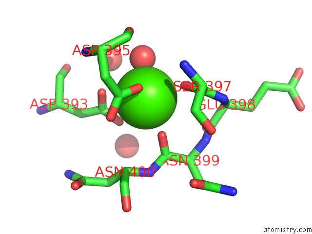
Mono view
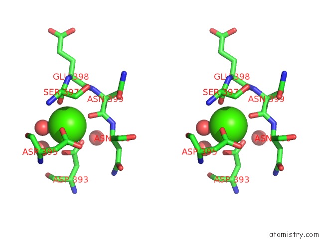
Stereo pair view
Mono view
Stereo pair view
A full contact list of Calcium with other atoms in the Ca binding
site number 1 of Crystal Structure of Tetrameric Fibrinogen-Like Recognition Domain of FIBCD1 with NEU5AC Ligand Bound within 5.0Å range:
|
Calcium binding site 2 out of 2 in 6zqy
Go back to
Calcium binding site 2 out
of 2 in the Crystal Structure of Tetrameric Fibrinogen-Like Recognition Domain of FIBCD1 with NEU5AC Ligand Bound
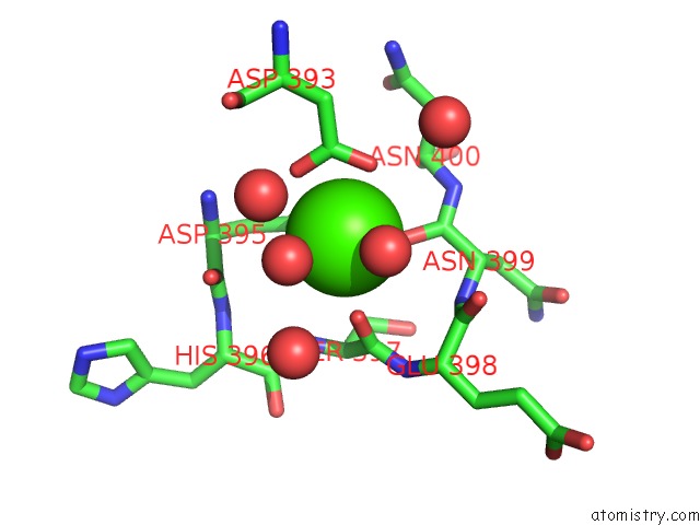
Mono view
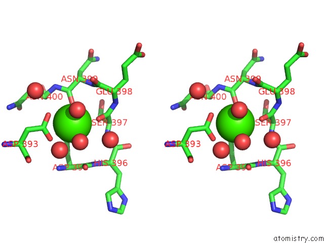
Stereo pair view
Mono view
Stereo pair view
A full contact list of Calcium with other atoms in the Ca binding
site number 2 of Crystal Structure of Tetrameric Fibrinogen-Like Recognition Domain of FIBCD1 with NEU5AC Ligand Bound within 5.0Å range:
|
Reference:
H.M.Williams,
J.B.Moeller,
I.Burns,
A.Schlosser,
G.L.Sorensen,
T.J.Greenhough,
U.Holmskov,
A.K.Shrive.
Crystal Structures of Human Immune Protein FIBCD1 Reveal An Extended Ligand Binding Site Compatible with Recognition of Chitin Oligomers To Be Published.
Page generated: Wed Jul 9 20:36:42 2025
Last articles
Mg in 7ELRMg in 7EMJ
Mg in 7ELS
Mg in 7ELP
Mg in 7ELQ
Mg in 7EJW
Mg in 7EL4
Mg in 7EKL
Mg in 7EJU
Mg in 7EJJ