Calcium »
PDB 6zr0-7aq1 »
7a1r »
Calcium in PDB 7a1r: Crystal Structure of the C2B Domain of Trypanosoma Brucei Extended Synaptotagmin (E-Syt)
Protein crystallography data
The structure of Crystal Structure of the C2B Domain of Trypanosoma Brucei Extended Synaptotagmin (E-Syt), PDB code: 7a1r
was solved by
G.Dong,
with X-Ray Crystallography technique. A brief refinement statistics is given in the table below:
| Resolution Low / High (Å) | 19.86 / 1.50 |
| Space group | P 21 21 21 |
| Cell size a, b, c (Å), α, β, γ (°) | 54.908, 57.532, 84.57, 90, 90, 90 |
| R / Rfree (%) | 17 / 19.1 |
Other elements in 7a1r:
The structure of Crystal Structure of the C2B Domain of Trypanosoma Brucei Extended Synaptotagmin (E-Syt) also contains other interesting chemical elements:
| Chlorine | (Cl) | 7 atoms |
| Sodium | (Na) | 1 atom |
Calcium Binding Sites:
The binding sites of Calcium atom in the Crystal Structure of the C2B Domain of Trypanosoma Brucei Extended Synaptotagmin (E-Syt)
(pdb code 7a1r). This binding sites where shown within
5.0 Angstroms radius around Calcium atom.
In total 4 binding sites of Calcium where determined in the Crystal Structure of the C2B Domain of Trypanosoma Brucei Extended Synaptotagmin (E-Syt), PDB code: 7a1r:
Jump to Calcium binding site number: 1; 2; 3; 4;
In total 4 binding sites of Calcium where determined in the Crystal Structure of the C2B Domain of Trypanosoma Brucei Extended Synaptotagmin (E-Syt), PDB code: 7a1r:
Jump to Calcium binding site number: 1; 2; 3; 4;
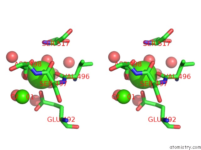
Calcium binding site 1 out of 4 in 7a1r
Go back to
Calcium binding site 1 out
of 4 in the Crystal Structure of the C2B Domain of Trypanosoma Brucei Extended Synaptotagmin (E-Syt)

Mono view

Stereo pair view
Mono view
Stereo pair view
A full contact list of Calcium with other atoms in the Ca binding
site number 1 of Crystal Structure of the C2B Domain of Trypanosoma Brucei Extended Synaptotagmin (E-Syt) within 5.0Å range:
|
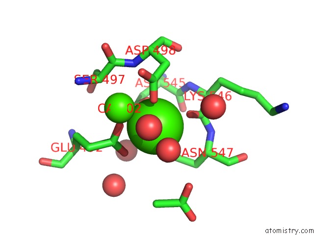
Calcium binding site 2 out of 4 in 7a1r
Go back to
Calcium binding site 2 out
of 4 in the Crystal Structure of the C2B Domain of Trypanosoma Brucei Extended Synaptotagmin (E-Syt)

Mono view
Stereo pair view
Mono view
Stereo pair view
A full contact list of Calcium with other atoms in the Ca binding
site number 2 of Crystal Structure of the C2B Domain of Trypanosoma Brucei Extended Synaptotagmin (E-Syt) within 5.0Å range:
|
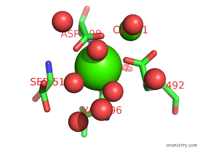
Calcium binding site 3 out of 4 in 7a1r
Go back to
Calcium binding site 3 out
of 4 in the Crystal Structure of the C2B Domain of Trypanosoma Brucei Extended Synaptotagmin (E-Syt)
Mono view

Stereo pair view
Mono view
Stereo pair view
A full contact list of Calcium with other atoms in the Ca binding
site number 3 of Crystal Structure of the C2B Domain of Trypanosoma Brucei Extended Synaptotagmin (E-Syt) within 5.0Å range:
|
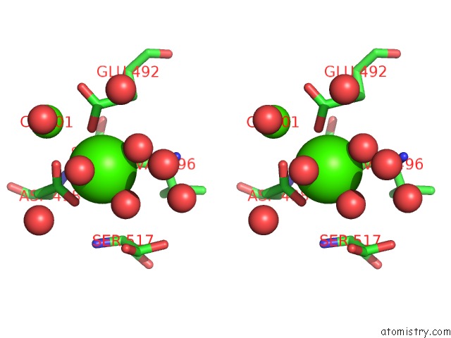
Calcium binding site 4 out of 4 in 7a1r
Go back to
Calcium binding site 4 out
of 4 in the Crystal Structure of the C2B Domain of Trypanosoma Brucei Extended Synaptotagmin (E-Syt)
Mono view
Stereo pair view
Mono view
Stereo pair view
A full contact list of Calcium with other atoms in the Ca binding
site number 4 of Crystal Structure of the C2B Domain of Trypanosoma Brucei Extended Synaptotagmin (E-Syt) within 5.0Å range:
|
Reference:
E.Stepinac,
N.Landrein,
D.Skwarzynska,
P.Wojcik,
J.Lesigang,
I.Lucic,
C.Y.He,
M.Bonhivers,
D.R.Robinson,
G.Dong.
Structural Studies of the Shortest Extended Synaptotagmin with Only Two C2 Domains From Trypanosoma Brucei . Iscience V. 24 02422 2021.
ISSN: ESSN 2589-0042
PubMed: 33997700
DOI: 10.1016/J.ISCI.2021.102422
Page generated: Wed Jul 9 20:39:53 2025
ISSN: ESSN 2589-0042
PubMed: 33997700
DOI: 10.1016/J.ISCI.2021.102422
Last articles
Fe in 4PH9Fe in 4PBU
Fe in 4PIX
Fe in 4PGK
Fe in 4PGI
Fe in 4PG1
Fe in 4PCU
Fe in 4PG0
Fe in 4P1B
Fe in 4P1C