Calcium »
PDB 7aq2-7b1l »
7azq »
Calcium in PDB 7azq: Crystal Structure of the Iron/Manganese Cambialistic Superoxide Dismutase From Rhodobacter Capsulatus Complex with Fe
Enzymatic activity of Crystal Structure of the Iron/Manganese Cambialistic Superoxide Dismutase From Rhodobacter Capsulatus Complex with Fe
All present enzymatic activity of Crystal Structure of the Iron/Manganese Cambialistic Superoxide Dismutase From Rhodobacter Capsulatus Complex with Fe:
1.15.1.1;
1.15.1.1;
Protein crystallography data
The structure of Crystal Structure of the Iron/Manganese Cambialistic Superoxide Dismutase From Rhodobacter Capsulatus Complex with Fe, PDB code: 7azq
was solved by
A.Ponce-Salvatierra,
J.A.Hermoso,
with X-Ray Crystallography technique. A brief refinement statistics is given in the table below:
| Resolution Low / High (Å) | 57.62 / 2.00 |
| Space group | P 41 21 2 |
| Cell size a, b, c (Å), α, β, γ (°) | 162.79, 162.79, 43.84, 90, 90, 90 |
| R / Rfree (%) | 16.9 / 18.8 |
Other elements in 7azq:
The structure of Crystal Structure of the Iron/Manganese Cambialistic Superoxide Dismutase From Rhodobacter Capsulatus Complex with Fe also contains other interesting chemical elements:
| Iron | (Fe) | 2 atoms |
Calcium Binding Sites:
The binding sites of Calcium atom in the Crystal Structure of the Iron/Manganese Cambialistic Superoxide Dismutase From Rhodobacter Capsulatus Complex with Fe
(pdb code 7azq). This binding sites where shown within
5.0 Angstroms radius around Calcium atom.
In total only one binding site of Calcium was determined in the Crystal Structure of the Iron/Manganese Cambialistic Superoxide Dismutase From Rhodobacter Capsulatus Complex with Fe, PDB code: 7azq:
In total only one binding site of Calcium was determined in the Crystal Structure of the Iron/Manganese Cambialistic Superoxide Dismutase From Rhodobacter Capsulatus Complex with Fe, PDB code: 7azq:
Calcium binding site 1 out of 1 in 7azq
Go back to
Calcium binding site 1 out
of 1 in the Crystal Structure of the Iron/Manganese Cambialistic Superoxide Dismutase From Rhodobacter Capsulatus Complex with Fe
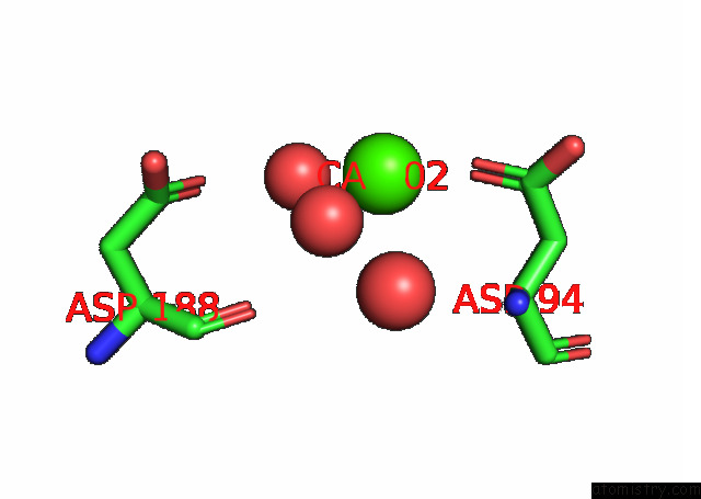
Mono view
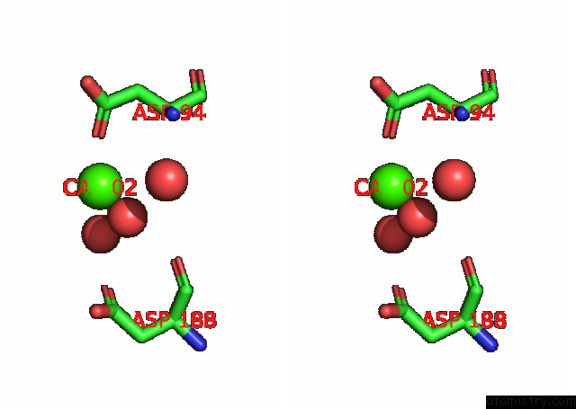
Stereo pair view
Mono view
Stereo pair view
A full contact list of Calcium with other atoms in the Ca binding
site number 1 of Crystal Structure of the Iron/Manganese Cambialistic Superoxide Dismutase From Rhodobacter Capsulatus Complex with Fe within 5.0Å range:
|
Reference:
A.Ponce-Salvatierra,
C.Di Capua,
J.Gonzalez,
N.Cortez,
J.A.Hermoso.
Structural and Functional Characterization of the Cambialistic Superoxide Dismutase From Rhodobacter Capsulatus. To Be Published.
Page generated: Wed Jul 9 20:54:02 2025
Last articles
Zn in 1X4WZn in 1X4V
Zn in 1X4U
Zn in 1X4S
Zn in 1X4L
Zn in 1X4K
Zn in 1X4J
Zn in 1X4I
Zn in 1X3H
Zn in 1X3Z