Calcium »
PDB 7c53-7cji »
7c74 »
Calcium in PDB 7c74: Crystal Structure of Yak Lactoperoxidase Using Data Obtained From Crystals Soaked in CACL2 at 2.73 A Resolution
Protein crystallography data
The structure of Crystal Structure of Yak Lactoperoxidase Using Data Obtained From Crystals Soaked in CACL2 at 2.73 A Resolution, PDB code: 7c74
was solved by
P.K.Singh,
V.Viswanathan,
S.N.Pandey,
N.Ahmad,
C.Rani,
P.Sharma,
P.Sharma,
T.P.Singh,
with X-Ray Crystallography technique. A brief refinement statistics is given in the table below:
| Resolution Low / High (Å) | 53.01 / 2.73 |
| Space group | P 1 21 1 |
| Cell size a, b, c (Å), α, β, γ (°) | 54.060, 80.590, 74.830, 90.00, 101.31, 90.00 |
| R / Rfree (%) | 19.5 / 29.2 |
Other elements in 7c74:
The structure of Crystal Structure of Yak Lactoperoxidase Using Data Obtained From Crystals Soaked in CACL2 at 2.73 A Resolution also contains other interesting chemical elements:
| Iron | (Fe) | 1 atom |
| Chlorine | (Cl) | 1 atom |
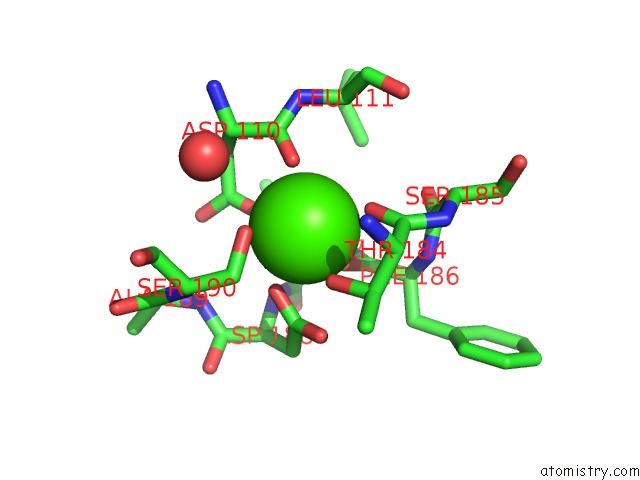
Calcium Binding Sites:
The binding sites of Calcium atom in the Crystal Structure of Yak Lactoperoxidase Using Data Obtained From Crystals Soaked in CACL2 at 2.73 A Resolution
(pdb code 7c74). This binding sites where shown within
5.0 Angstroms radius around Calcium atom.
In total only one binding site of Calcium was determined in the Crystal Structure of Yak Lactoperoxidase Using Data Obtained From Crystals Soaked in CACL2 at 2.73 A Resolution, PDB code: 7c74:
In total only one binding site of Calcium was determined in the Crystal Structure of Yak Lactoperoxidase Using Data Obtained From Crystals Soaked in CACL2 at 2.73 A Resolution, PDB code: 7c74:
Calcium binding site 1 out of 1 in 7c74
Go back to
Calcium binding site 1 out
of 1 in the Crystal Structure of Yak Lactoperoxidase Using Data Obtained From Crystals Soaked in CACL2 at 2.73 A Resolution
Mono view

Stereo pair view
Mono view
Stereo pair view
A full contact list of Calcium with other atoms in the Ca binding
site number 1 of Crystal Structure of Yak Lactoperoxidase Using Data Obtained From Crystals Soaked in CACL2 at 2.73 A Resolution within 5.0Å range:
|
Reference:
P.K.Singh,
V.Viswanathan,
S.N.Pandey,
N.Ahmad,
C.Rani,
P.Sharma,
S.Sharma,
T.P.Singh.
Crystal Structure of Yak Lactoperoxidase Using Data Obtained From Crystals Soaked in CACL2 at 2.73 A Resolution To Be Published.
Page generated: Wed Jul 9 21:09:03 2025
Last articles
Zn in 8PK7Zn in 8PIM
Zn in 8PJN
Zn in 8PIL
Zn in 8PID
Zn in 8PI5
Zn in 8PIB
Zn in 8PHK
Zn in 8PHL
Zn in 8PH9