Calcium »
PDB 7dlh-7e6q »
7e4r »
Calcium in PDB 7e4r: Crystal Structure of Tubulin in Complex with D-DM1-Sme
Protein crystallography data
The structure of Crystal Structure of Tubulin in Complex with D-DM1-Sme, PDB code: 7e4r
was solved by
Y.Wang,
W.Li,
with X-Ray Crystallography technique. A brief refinement statistics is given in the table below:
| Resolution Low / High (Å) | 45.45 / 2.60 |
| Space group | P 21 21 21 |
| Cell size a, b, c (Å), α, β, γ (°) | 104.982, 157.229, 182.574, 90, 90, 90 |
| R / Rfree (%) | 22.4 / 26.5 |
Other elements in 7e4r:
The structure of Crystal Structure of Tubulin in Complex with D-DM1-Sme also contains other interesting chemical elements:
| Magnesium | (Mg) | 4 atoms |
| Chlorine | (Cl) | 1 atom |
Calcium Binding Sites:
The binding sites of Calcium atom in the Crystal Structure of Tubulin in Complex with D-DM1-Sme
(pdb code 7e4r). This binding sites where shown within
5.0 Angstroms radius around Calcium atom.
In total 2 binding sites of Calcium where determined in the Crystal Structure of Tubulin in Complex with D-DM1-Sme, PDB code: 7e4r:
Jump to Calcium binding site number: 1; 2;
In total 2 binding sites of Calcium where determined in the Crystal Structure of Tubulin in Complex with D-DM1-Sme, PDB code: 7e4r:
Jump to Calcium binding site number: 1; 2;
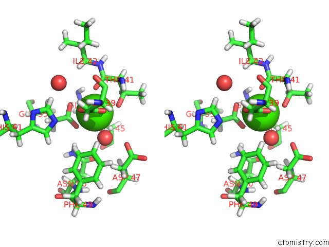
Calcium binding site 1 out of 2 in 7e4r
Go back to
Calcium binding site 1 out
of 2 in the Crystal Structure of Tubulin in Complex with D-DM1-Sme
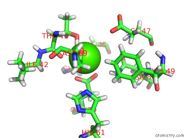
Mono view

Stereo pair view
Mono view
Stereo pair view
A full contact list of Calcium with other atoms in the Ca binding
site number 1 of Crystal Structure of Tubulin in Complex with D-DM1-Sme within 5.0Å range:
|
Calcium binding site 2 out of 2 in 7e4r
Go back to
Calcium binding site 2 out
of 2 in the Crystal Structure of Tubulin in Complex with D-DM1-Sme
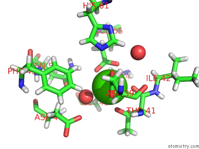
Mono view
Stereo pair view
Mono view
Stereo pair view
A full contact list of Calcium with other atoms in the Ca binding
site number 2 of Crystal Structure of Tubulin in Complex with D-DM1-Sme within 5.0Å range:
|
Reference:
W.Li,
M.Huang,
Y.Li,
A.Xia,
L.Tan,
Z.Zhang,
Y.Wang,
J.Yang.
C3 Ester Side Chain Plays A Pivotal Role in the Antitumor Activity of Maytansinoids. Biochem.Biophys.Res.Commun. V. 566 197 2021.
ISSN: ESSN 1090-2104
PubMed: 34144258
DOI: 10.1016/J.BBRC.2021.05.071
Page generated: Wed Jul 9 21:45:05 2025
ISSN: ESSN 1090-2104
PubMed: 34144258
DOI: 10.1016/J.BBRC.2021.05.071
Last articles
Mg in 4DUXMg in 4DUW
Mg in 4DUV
Mg in 4DUO
Mg in 4DUG
Mg in 4DTY
Mg in 4DTW
Mg in 4DTH
Mg in 4DTF
Mg in 4DSU