Calcium »
PDB 7e6t-7et8 »
7ea4 »
Calcium in PDB 7ea4: Crystal Structure of L182E D-Succinylase (Dsa) From Cupriavidus Sp. P4-10-C
Protein crystallography data
The structure of Crystal Structure of L182E D-Succinylase (Dsa) From Cupriavidus Sp. P4-10-C, PDB code: 7ea4
was solved by
M.Yamasaki,
Y.Sumida,
with X-Ray Crystallography technique. A brief refinement statistics is given in the table below:
| Resolution Low / High (Å) | 42.93 / 1.95 |
| Space group | P 21 21 21 |
| Cell size a, b, c (Å), α, β, γ (°) | 62.057, 129.806, 200.68, 90, 90, 90 |
| R / Rfree (%) | 16.4 / 20.8 |
Other elements in 7ea4:
The structure of Crystal Structure of L182E D-Succinylase (Dsa) From Cupriavidus Sp. P4-10-C also contains other interesting chemical elements:
| Chlorine | (Cl) | 2 atoms |
| Arsenic | (As) | 2 atoms |
Calcium Binding Sites:
The binding sites of Calcium atom in the Crystal Structure of L182E D-Succinylase (Dsa) From Cupriavidus Sp. P4-10-C
(pdb code 7ea4). This binding sites where shown within
5.0 Angstroms radius around Calcium atom.
In total 2 binding sites of Calcium where determined in the Crystal Structure of L182E D-Succinylase (Dsa) From Cupriavidus Sp. P4-10-C, PDB code: 7ea4:
Jump to Calcium binding site number: 1; 2;
In total 2 binding sites of Calcium where determined in the Crystal Structure of L182E D-Succinylase (Dsa) From Cupriavidus Sp. P4-10-C, PDB code: 7ea4:
Jump to Calcium binding site number: 1; 2;
Calcium binding site 1 out of 2 in 7ea4
Go back to
Calcium binding site 1 out
of 2 in the Crystal Structure of L182E D-Succinylase (Dsa) From Cupriavidus Sp. P4-10-C
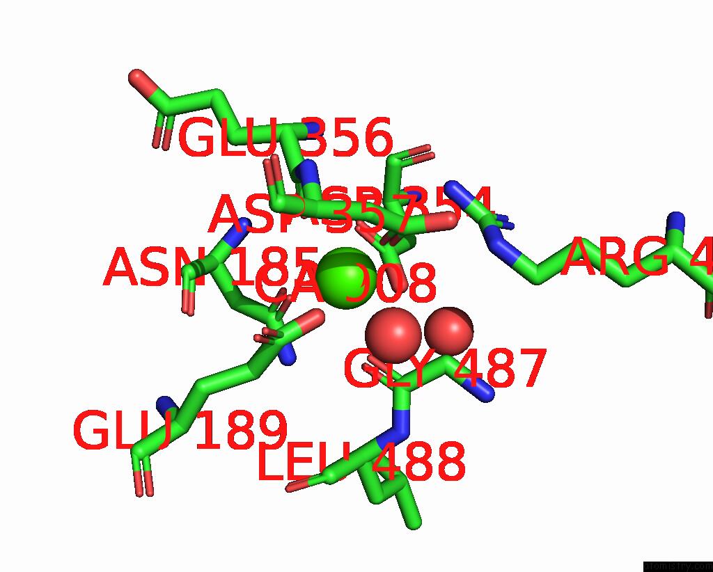
Mono view
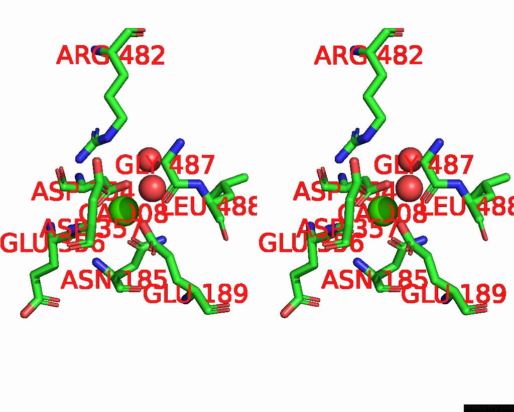
Stereo pair view
Mono view
Stereo pair view
A full contact list of Calcium with other atoms in the Ca binding
site number 1 of Crystal Structure of L182E D-Succinylase (Dsa) From Cupriavidus Sp. P4-10-C within 5.0Å range:
|
Calcium binding site 2 out of 2 in 7ea4
Go back to
Calcium binding site 2 out
of 2 in the Crystal Structure of L182E D-Succinylase (Dsa) From Cupriavidus Sp. P4-10-C
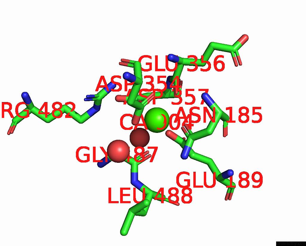
Mono view
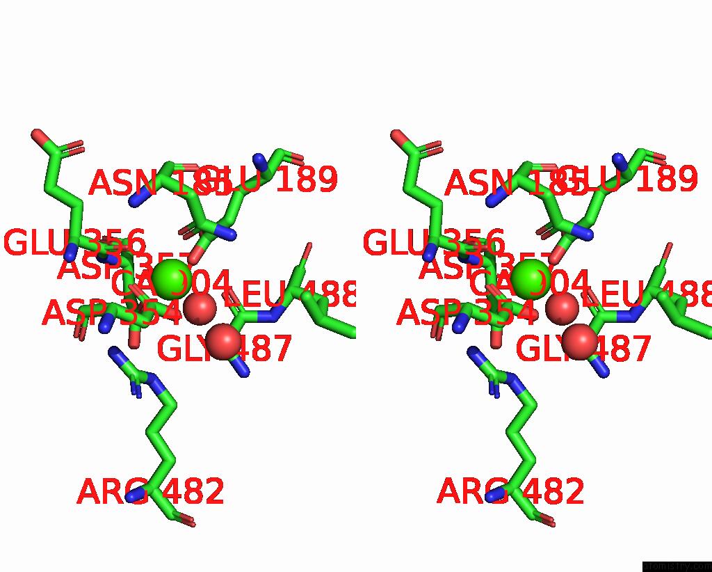
Stereo pair view
Mono view
Stereo pair view
A full contact list of Calcium with other atoms in the Ca binding
site number 2 of Crystal Structure of L182E D-Succinylase (Dsa) From Cupriavidus Sp. P4-10-C within 5.0Å range:
|
Reference:
Y.Sumida,
M.Yamasaki,
Y.Nishiya,
S.Kumagai,
T.Yamada,
M.Azuma.
Protein Engineering of D-Succinylase From Cupriavidus Sp. For D-Amino Acid Synthesis and the Structural Implications. Adv.Synth.Catal. V. 363 4770 2021.
ISSN: ESSN 1615-4169
DOI: 10.1002/ADSC.202100587
Page generated: Wed Jul 9 21:47:38 2025
ISSN: ESSN 1615-4169
DOI: 10.1002/ADSC.202100587
Last articles
K in 3F2QK in 3EZT
K in 3F06
K in 3EZP
K in 3EXH
K in 3EWF
K in 3EXI
K in 3EXF
K in 3EXE
K in 3EUI