Calcium »
PDB 7e6t-7et8 »
7esi »
Calcium in PDB 7esi: Crystal Structure of the Collagenase Unit of A Vibrio Collagenase From Vibrio Harveyi VHJR7 at 1. 8 Angstrom Resolution.
Protein crystallography data
The structure of Crystal Structure of the Collagenase Unit of A Vibrio Collagenase From Vibrio Harveyi VHJR7 at 1. 8 Angstrom Resolution., PDB code: 7esi
was solved by
H.Y.Cao,
Y.Wang,
M.Peng,
Y.Z.Zhang,
with X-Ray Crystallography technique. A brief refinement statistics is given in the table below:
| Resolution Low / High (Å) | 49.79 / 1.80 |
| Space group | P 1 21 1 |
| Cell size a, b, c (Å), α, β, γ (°) | 74.444, 50.068, 90.681, 90, 104.72, 90 |
| R / Rfree (%) | 15.9 / 18.7 |
Other elements in 7esi:
The structure of Crystal Structure of the Collagenase Unit of A Vibrio Collagenase From Vibrio Harveyi VHJR7 at 1. 8 Angstrom Resolution. also contains other interesting chemical elements:
| Zinc | (Zn) | 1 atom |
Calcium Binding Sites:
The binding sites of Calcium atom in the Crystal Structure of the Collagenase Unit of A Vibrio Collagenase From Vibrio Harveyi VHJR7 at 1. 8 Angstrom Resolution.
(pdb code 7esi). This binding sites where shown within
5.0 Angstroms radius around Calcium atom.
In total only one binding site of Calcium was determined in the Crystal Structure of the Collagenase Unit of A Vibrio Collagenase From Vibrio Harveyi VHJR7 at 1. 8 Angstrom Resolution., PDB code: 7esi:
In total only one binding site of Calcium was determined in the Crystal Structure of the Collagenase Unit of A Vibrio Collagenase From Vibrio Harveyi VHJR7 at 1. 8 Angstrom Resolution., PDB code: 7esi:
Calcium binding site 1 out of 1 in 7esi
Go back to
Calcium binding site 1 out
of 1 in the Crystal Structure of the Collagenase Unit of A Vibrio Collagenase From Vibrio Harveyi VHJR7 at 1. 8 Angstrom Resolution.
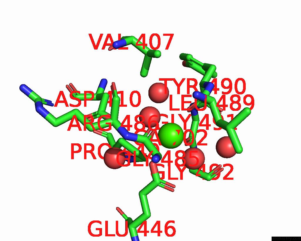
Mono view
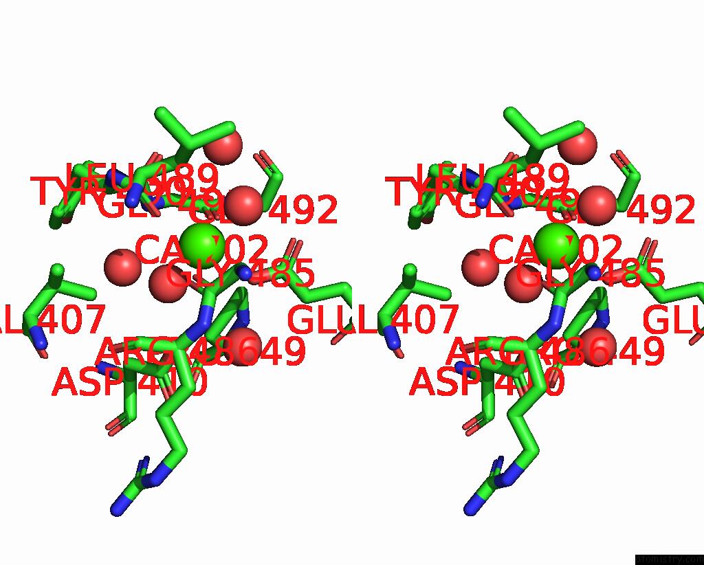
Stereo pair view
Mono view
Stereo pair view
A full contact list of Calcium with other atoms in the Ca binding
site number 1 of Crystal Structure of the Collagenase Unit of A Vibrio Collagenase From Vibrio Harveyi VHJR7 at 1. 8 Angstrom Resolution. within 5.0Å range:
|
Reference:
Y.Wang,
H.Y.Cao,
M.Peng.
Crystal Structure of the Collagenase Unit of A Vibrio Collagenase From Vibrio Harveyi VHJR7 at 1. 8 Angstrom Resolution. To Be Published.
Page generated: Wed Jul 9 21:54:42 2025
Last articles
Mg in 8WGHMg in 8WMO
Mg in 8WMN
Mg in 8WMM
Mg in 8WEY
Mg in 8WKG
Mg in 8WKF
Mg in 8WIM
Mg in 8WIL
Mg in 8WH2