Calcium »
PDB 7jlu-7k04 »
7jmr »
Calcium in PDB 7jmr: Crystal Structure of the Pea Pathogenicity Protein 2 From Madurella Mycetomatis
Protein crystallography data
The structure of Crystal Structure of the Pea Pathogenicity Protein 2 From Madurella Mycetomatis, PDB code: 7jmr
was solved by
M.Zeug,
N.Markovic,
C.V.Iancu,
J.Tripp,
M.Oreb,
J.Choe,
with X-Ray Crystallography technique. A brief refinement statistics is given in the table below:
| Resolution Low / High (Å) | 38.74 / 1.67 |
| Space group | P 3 2 1 |
| Cell size a, b, c (Å), α, β, γ (°) | 98.853, 98.853, 62.38, 90, 90, 120 |
| R / Rfree (%) | 17.7 / 19.6 |
Other elements in 7jmr:
The structure of Crystal Structure of the Pea Pathogenicity Protein 2 From Madurella Mycetomatis also contains other interesting chemical elements:
| Potassium | (K) | 1 atom |
Calcium Binding Sites:
The binding sites of Calcium atom in the Crystal Structure of the Pea Pathogenicity Protein 2 From Madurella Mycetomatis
(pdb code 7jmr). This binding sites where shown within
5.0 Angstroms radius around Calcium atom.
In total 3 binding sites of Calcium where determined in the Crystal Structure of the Pea Pathogenicity Protein 2 From Madurella Mycetomatis, PDB code: 7jmr:
Jump to Calcium binding site number: 1; 2; 3;
In total 3 binding sites of Calcium where determined in the Crystal Structure of the Pea Pathogenicity Protein 2 From Madurella Mycetomatis, PDB code: 7jmr:
Jump to Calcium binding site number: 1; 2; 3;
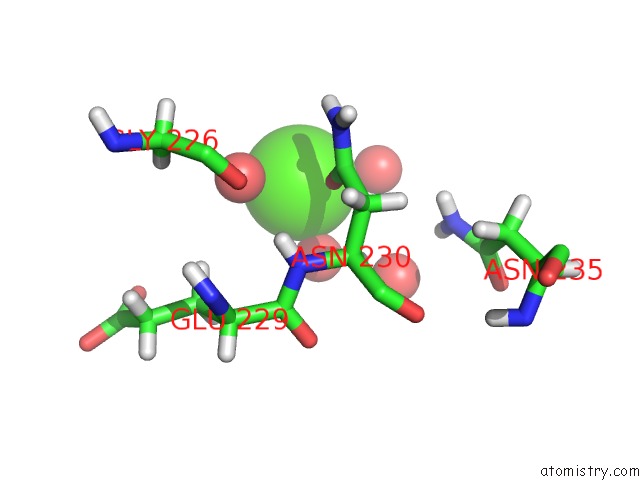
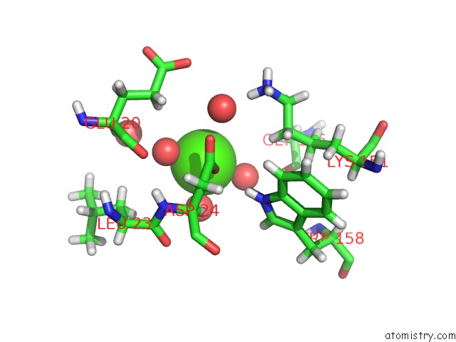
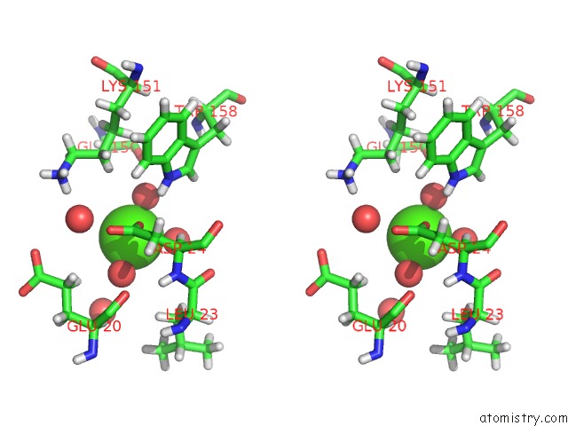
Calcium binding site 1 out of 3 in 7jmr
Go back to
Calcium binding site 1 out
of 3 in the Crystal Structure of the Pea Pathogenicity Protein 2 From Madurella Mycetomatis
Mono view
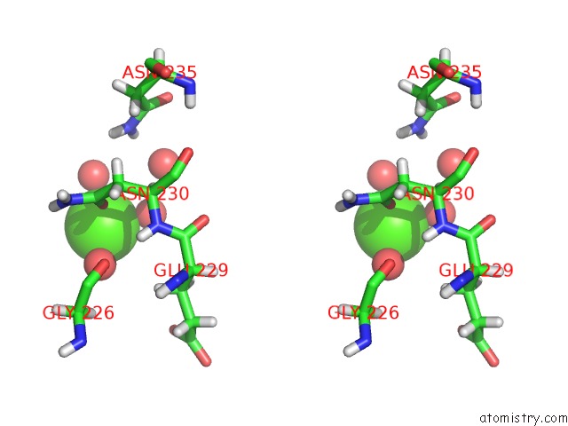
Stereo pair view
Mono view
Stereo pair view
A full contact list of Calcium with other atoms in the Ca binding
site number 1 of Crystal Structure of the Pea Pathogenicity Protein 2 From Madurella Mycetomatis within 5.0Å range:
|
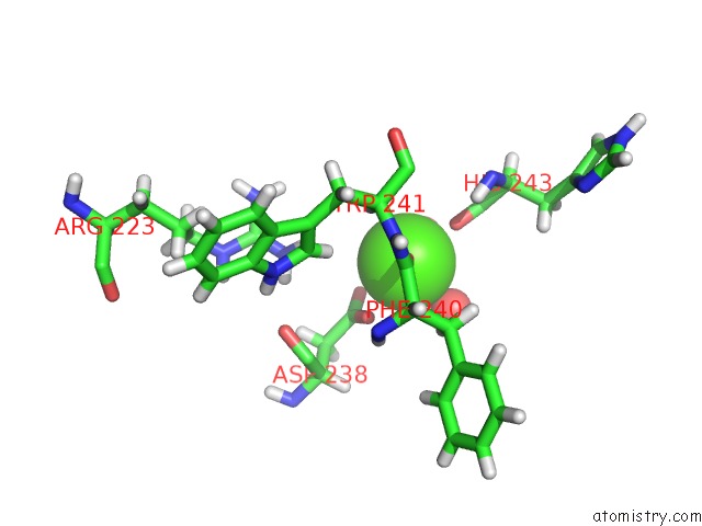
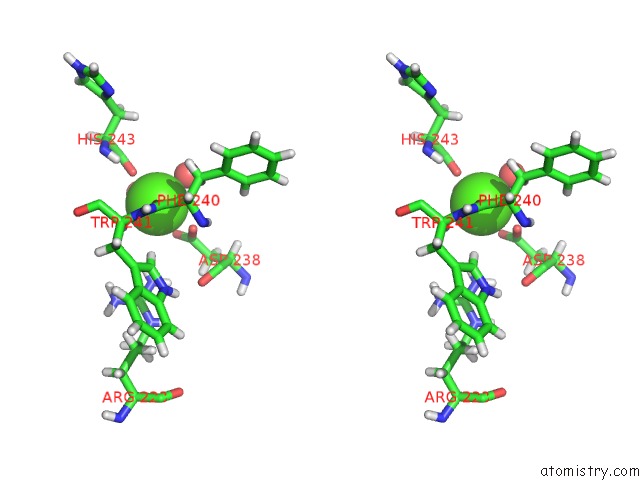
Calcium binding site 2 out of 3 in 7jmr
Go back to
Calcium binding site 2 out
of 3 in the Crystal Structure of the Pea Pathogenicity Protein 2 From Madurella Mycetomatis
Mono view
Stereo pair view
Mono view
Stereo pair view
A full contact list of Calcium with other atoms in the Ca binding
site number 2 of Crystal Structure of the Pea Pathogenicity Protein 2 From Madurella Mycetomatis within 5.0Å range:
|
Calcium binding site 3 out of 3 in 7jmr
Go back to
Calcium binding site 3 out
of 3 in the Crystal Structure of the Pea Pathogenicity Protein 2 From Madurella Mycetomatis
Mono view
Stereo pair view
Mono view
Stereo pair view
A full contact list of Calcium with other atoms in the Ca binding
site number 3 of Crystal Structure of the Pea Pathogenicity Protein 2 From Madurella Mycetomatis within 5.0Å range:
|
Reference:
M.Zeug,
N.Markovic,
C.V.Iancu,
J.Tripp,
M.Oreb,
J.Y.Choe.
Crystal Structures of Non-Oxidative Decarboxylases Reveal A New Mechanism of Action with A Catalytic Dyad and Structural Twists. Sci Rep V. 11 3056 2021.
ISSN: ESSN 2045-2322
PubMed: 33542397
DOI: 10.1038/S41598-021-82660-Z
Page generated: Wed Jul 9 22:43:20 2025
ISSN: ESSN 2045-2322
PubMed: 33542397
DOI: 10.1038/S41598-021-82660-Z
Last articles
Fe in 2YXOFe in 2YRS
Fe in 2YXC
Fe in 2YNM
Fe in 2YVJ
Fe in 2YP1
Fe in 2YU2
Fe in 2YU1
Fe in 2YQB
Fe in 2YOO