Calcium »
PDB 7jlu-7k04 »
7jmv »
Calcium in PDB 7jmv: Crystal Structure of the Pea Pathogenicity Protein 2 From Madurella Mycetomatis Complexed with 4-Nitrocatechol
Protein crystallography data
The structure of Crystal Structure of the Pea Pathogenicity Protein 2 From Madurella Mycetomatis Complexed with 4-Nitrocatechol, PDB code: 7jmv
was solved by
M.Zeug,
N.Markovic,
C.V.Iancu,
J.Tripp,
M.Oreb,
J.Choe,
with X-Ray Crystallography technique. A brief refinement statistics is given in the table below:
| Resolution Low / High (Å) | 42.76 / 1.57 |
| Space group | P 3 2 1 |
| Cell size a, b, c (Å), α, β, γ (°) | 98.74, 98.74, 62.287, 90, 90, 120 |
| R / Rfree (%) | 17.7 / 19.1 |
Other elements in 7jmv:
The structure of Crystal Structure of the Pea Pathogenicity Protein 2 From Madurella Mycetomatis Complexed with 4-Nitrocatechol also contains other interesting chemical elements:
| Potassium | (K) | 1 atom |
Calcium Binding Sites:
The binding sites of Calcium atom in the Crystal Structure of the Pea Pathogenicity Protein 2 From Madurella Mycetomatis Complexed with 4-Nitrocatechol
(pdb code 7jmv). This binding sites where shown within
5.0 Angstroms radius around Calcium atom.
In total 2 binding sites of Calcium where determined in the Crystal Structure of the Pea Pathogenicity Protein 2 From Madurella Mycetomatis Complexed with 4-Nitrocatechol, PDB code: 7jmv:
Jump to Calcium binding site number: 1; 2;
In total 2 binding sites of Calcium where determined in the Crystal Structure of the Pea Pathogenicity Protein 2 From Madurella Mycetomatis Complexed with 4-Nitrocatechol, PDB code: 7jmv:
Jump to Calcium binding site number: 1; 2;
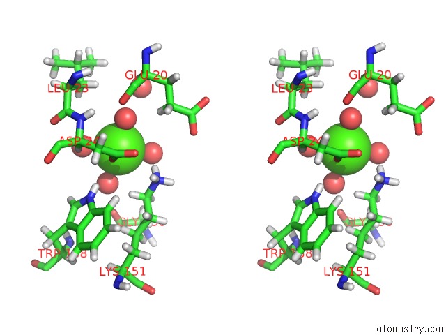
Calcium binding site 1 out of 2 in 7jmv
Go back to
Calcium binding site 1 out
of 2 in the Crystal Structure of the Pea Pathogenicity Protein 2 From Madurella Mycetomatis Complexed with 4-Nitrocatechol

Mono view

Stereo pair view
Mono view
Stereo pair view
A full contact list of Calcium with other atoms in the Ca binding
site number 1 of Crystal Structure of the Pea Pathogenicity Protein 2 From Madurella Mycetomatis Complexed with 4-Nitrocatechol within 5.0Å range:
|
Calcium binding site 2 out of 2 in 7jmv
Go back to
Calcium binding site 2 out
of 2 in the Crystal Structure of the Pea Pathogenicity Protein 2 From Madurella Mycetomatis Complexed with 4-Nitrocatechol

Mono view
Stereo pair view
Mono view
Stereo pair view
A full contact list of Calcium with other atoms in the Ca binding
site number 2 of Crystal Structure of the Pea Pathogenicity Protein 2 From Madurella Mycetomatis Complexed with 4-Nitrocatechol within 5.0Å range:
|
Reference:
M.Zeug,
N.Markovic,
C.V.Iancu,
J.Tripp,
M.Oreb,
J.Y.Choe.
Crystal Structures of Non-Oxidative Decarboxylases Reveal A New Mechanism of Action with A Catalytic Dyad and Structural Twists. Sci Rep V. 11 3056 2021.
ISSN: ESSN 2045-2322
PubMed: 33542397
DOI: 10.1038/S41598-021-82660-Z
Page generated: Wed Jul 9 22:43:32 2025
ISSN: ESSN 2045-2322
PubMed: 33542397
DOI: 10.1038/S41598-021-82660-Z
Last articles
Br in 9R0QBr in 9J73
Br in 9BJ5
Br in 8Y72
Au in 9D33
As in 9O9I
Al in 9GSG
Zr in 1XC1
Zr in 6Y7P
Zr in 6GNL