Calcium »
PDB 7jlu-7k04 »
7jty »
Calcium in PDB 7jty: Co-Crystal Structure of Alpha Glucosidase with Compound 1
Protein crystallography data
The structure of Co-Crystal Structure of Alpha Glucosidase with Compound 1, PDB code: 7jty
was solved by
S.S.Karade,
R.A.Mariuzza,
with X-Ray Crystallography technique. A brief refinement statistics is given in the table below:
| Resolution Low / High (Å) | 42.17 / 2.21 |
| Space group | P 32 |
| Cell size a, b, c (Å), α, β, γ (°) | 102.561, 102.561, 239.559, 90, 90, 120 |
| R / Rfree (%) | 17 / 20.3 |
Calcium Binding Sites:
The binding sites of Calcium atom in the Co-Crystal Structure of Alpha Glucosidase with Compound 1
(pdb code 7jty). This binding sites where shown within
5.0 Angstroms radius around Calcium atom.
In total 4 binding sites of Calcium where determined in the Co-Crystal Structure of Alpha Glucosidase with Compound 1, PDB code: 7jty:
Jump to Calcium binding site number: 1; 2; 3; 4;
In total 4 binding sites of Calcium where determined in the Co-Crystal Structure of Alpha Glucosidase with Compound 1, PDB code: 7jty:
Jump to Calcium binding site number: 1; 2; 3; 4;
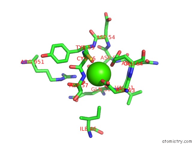
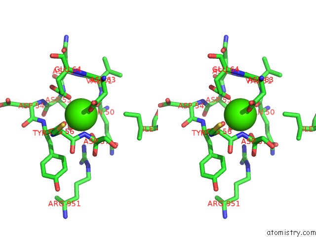
Calcium binding site 1 out of 4 in 7jty
Go back to
Calcium binding site 1 out
of 4 in the Co-Crystal Structure of Alpha Glucosidase with Compound 1
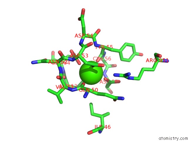
Mono view
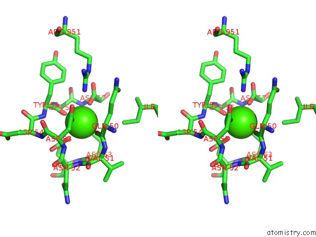
Stereo pair view
Mono view
Stereo pair view
A full contact list of Calcium with other atoms in the Ca binding
site number 1 of Co-Crystal Structure of Alpha Glucosidase with Compound 1 within 5.0Å range:
|
Calcium binding site 2 out of 4 in 7jty
Go back to
Calcium binding site 2 out
of 4 in the Co-Crystal Structure of Alpha Glucosidase with Compound 1
Mono view
Stereo pair view
Mono view
Stereo pair view
A full contact list of Calcium with other atoms in the Ca binding
site number 2 of Co-Crystal Structure of Alpha Glucosidase with Compound 1 within 5.0Å range:
|
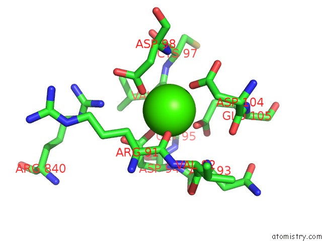
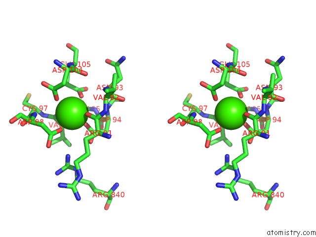
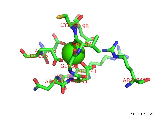
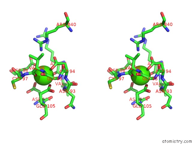
Calcium binding site 3 out of 4 in 7jty
Go back to
Calcium binding site 3 out
of 4 in the Co-Crystal Structure of Alpha Glucosidase with Compound 1
Mono view
Stereo pair view
Mono view
Stereo pair view
A full contact list of Calcium with other atoms in the Ca binding
site number 3 of Co-Crystal Structure of Alpha Glucosidase with Compound 1 within 5.0Å range:
|
Calcium binding site 4 out of 4 in 7jty
Go back to
Calcium binding site 4 out
of 4 in the Co-Crystal Structure of Alpha Glucosidase with Compound 1
Mono view
Stereo pair view
Mono view
Stereo pair view
A full contact list of Calcium with other atoms in the Ca binding
site number 4 of Co-Crystal Structure of Alpha Glucosidase with Compound 1 within 5.0Å range:
|
Reference:
S.S.Karade,
R.A.Mariuzza.
Co-Crystal Structure of Mouse Alpha Glucosidase (N97D) with Compound 1 To Be Published.
Page generated: Wed Jul 9 22:46:03 2025
Last articles
Gd in 7TSOGd in 7TSN
Gd in 7TSM
Gd in 7TSL
Gd in 7TSK
Gd in 7TSI
Gd in 7TSH
Gd in 7TSG
Gd in 7PH7
Gd in 7PH4