Calcium »
PDB 7or8-7p9r »
7p1h »
Calcium in PDB 7p1h: Structure of the V. Vulnificus Exoy-G-Actin-Profilin Complex
Calcium Binding Sites:
The binding sites of Calcium atom in the Structure of the V. Vulnificus Exoy-G-Actin-Profilin Complex
(pdb code 7p1h). This binding sites where shown within
5.0 Angstroms radius around Calcium atom.
In total only one binding site of Calcium was determined in the Structure of the V. Vulnificus Exoy-G-Actin-Profilin Complex, PDB code: 7p1h:
In total only one binding site of Calcium was determined in the Structure of the V. Vulnificus Exoy-G-Actin-Profilin Complex, PDB code: 7p1h:
Calcium binding site 1 out of 1 in 7p1h
Go back to
Calcium binding site 1 out
of 1 in the Structure of the V. Vulnificus Exoy-G-Actin-Profilin Complex
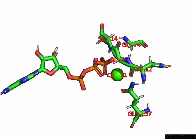
Mono view
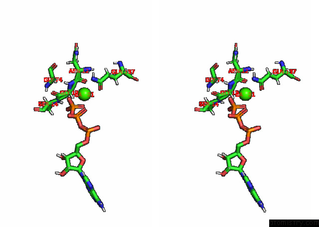
Stereo pair view
Mono view
Stereo pair view
A full contact list of Calcium with other atoms in the Ca binding
site number 1 of Structure of the V. Vulnificus Exoy-G-Actin-Profilin Complex within 5.0Å range:
|
Reference:
A.Belyy,
F.Merino,
U.Mechold,
S.Raunser.
Mechanism of Actin-Dependent Activation of Nucleotidyl Cyclase Toxins From Bacterial Human Pathogens To Be Published.
DOI: 10.1038/S41467-021-26889-2
Page generated: Wed Jul 9 23:59:19 2025
DOI: 10.1038/S41467-021-26889-2
Last articles
Mg in 6CA4Mg in 6C90
Mg in 6CA0
Mg in 6C9Y
Mg in 6C8Z
Mg in 6C8P
Mg in 6C8N
Mg in 6C8O
Mg in 6C8D
Mg in 6C8L