Calcium »
PDB 7s6e-7skl »
7sie »
Calcium in PDB 7sie: Structure of Aap A-Domain (Residues 351-605) From Staphylococcus Epidermidis
Protein crystallography data
The structure of Structure of Aap A-Domain (Residues 351-605) From Staphylococcus Epidermidis, PDB code: 7sie
was solved by
K.E.Atkin,
A.S.Brentnall,
E.J.Dodson,
F.Whelan,
L.Clark,
J.P.Turkenburg,
J.R.Potts,
with X-Ray Crystallography technique. A brief refinement statistics is given in the table below:
| Resolution Low / High (Å) | 16.53 / 1.30 |
| Space group | P 21 21 21 |
| Cell size a, b, c (Å), α, β, γ (°) | 35.853, 68.693, 109.198, 90, 90, 90 |
| R / Rfree (%) | 10 / 12.4 |
Other elements in 7sie:
The structure of Structure of Aap A-Domain (Residues 351-605) From Staphylococcus Epidermidis also contains other interesting chemical elements:
| Chlorine | (Cl) | 1 atom |
Calcium Binding Sites:
The binding sites of Calcium atom in the Structure of Aap A-Domain (Residues 351-605) From Staphylococcus Epidermidis
(pdb code 7sie). This binding sites where shown within
5.0 Angstroms radius around Calcium atom.
In total only one binding site of Calcium was determined in the Structure of Aap A-Domain (Residues 351-605) From Staphylococcus Epidermidis, PDB code: 7sie:
In total only one binding site of Calcium was determined in the Structure of Aap A-Domain (Residues 351-605) From Staphylococcus Epidermidis, PDB code: 7sie:
Calcium binding site 1 out of 1 in 7sie
Go back to
Calcium binding site 1 out
of 1 in the Structure of Aap A-Domain (Residues 351-605) From Staphylococcus Epidermidis
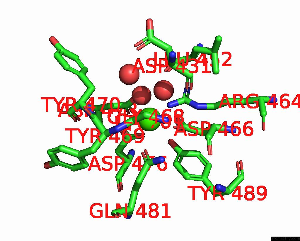
Mono view
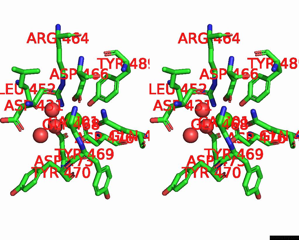
Stereo pair view
Mono view
Stereo pair view
A full contact list of Calcium with other atoms in the Ca binding
site number 1 of Structure of Aap A-Domain (Residues 351-605) From Staphylococcus Epidermidis within 5.0Å range:
|
Reference:
K.E.Atkin,
A.S.Brentnall,
L.Clark,
F.Whelan,
J.P.Turkenburg,
J.R.Potts.
Structure of Aap A-Domain (Residues 351-605) From Staphylococcus Epidermidis To Be Published.
Page generated: Thu Jul 10 01:00:26 2025
Last articles
Mg in 6MXTMg in 6MXC
Mg in 6MXO
Mg in 6MXG
Mg in 6MXD
Mg in 6MXB
Mg in 6MW7
Mg in 6MWK
Mg in 6MTI
Mg in 6MVE