Calcium »
PDB 7vmk-7wa0 »
7w4z »
Calcium in PDB 7w4z: Crystal Structure of Fragmin Domain-1 in Complex with Actin (Amppnp- Form)
Protein crystallography data
The structure of Crystal Structure of Fragmin Domain-1 in Complex with Actin (Amppnp- Form), PDB code: 7w4z
was solved by
S.Takeda,
with X-Ray Crystallography technique. A brief refinement statistics is given in the table below:
| Resolution Low / High (Å) | 29.35 / 1.15 |
| Space group | P 21 21 21 |
| Cell size a, b, c (Å), α, β, γ (°) | 57.09, 91.07, 114.85, 90, 90, 90 |
| R / Rfree (%) | 14.3 / 15.8 |
Other elements in 7w4z:
The structure of Crystal Structure of Fragmin Domain-1 in Complex with Actin (Amppnp- Form) also contains other interesting chemical elements:
| Magnesium | (Mg) | 1 atom |
Calcium Binding Sites:
The binding sites of Calcium atom in the Crystal Structure of Fragmin Domain-1 in Complex with Actin (Amppnp- Form)
(pdb code 7w4z). This binding sites where shown within
5.0 Angstroms radius around Calcium atom.
In total 2 binding sites of Calcium where determined in the Crystal Structure of Fragmin Domain-1 in Complex with Actin (Amppnp- Form), PDB code: 7w4z:
Jump to Calcium binding site number: 1; 2;
In total 2 binding sites of Calcium where determined in the Crystal Structure of Fragmin Domain-1 in Complex with Actin (Amppnp- Form), PDB code: 7w4z:
Jump to Calcium binding site number: 1; 2;
Calcium binding site 1 out of 2 in 7w4z
Go back to
Calcium binding site 1 out
of 2 in the Crystal Structure of Fragmin Domain-1 in Complex with Actin (Amppnp- Form)
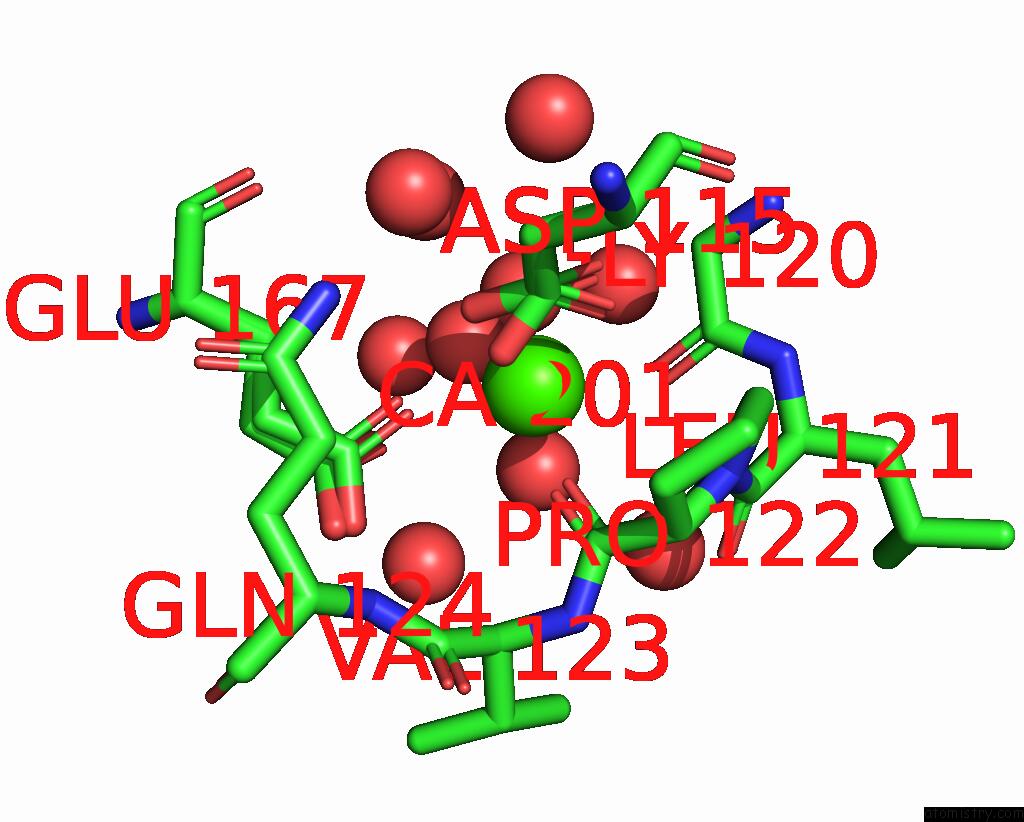
Mono view

Stereo pair view
Mono view
Stereo pair view
A full contact list of Calcium with other atoms in the Ca binding
site number 1 of Crystal Structure of Fragmin Domain-1 in Complex with Actin (Amppnp- Form) within 5.0Å range:
|
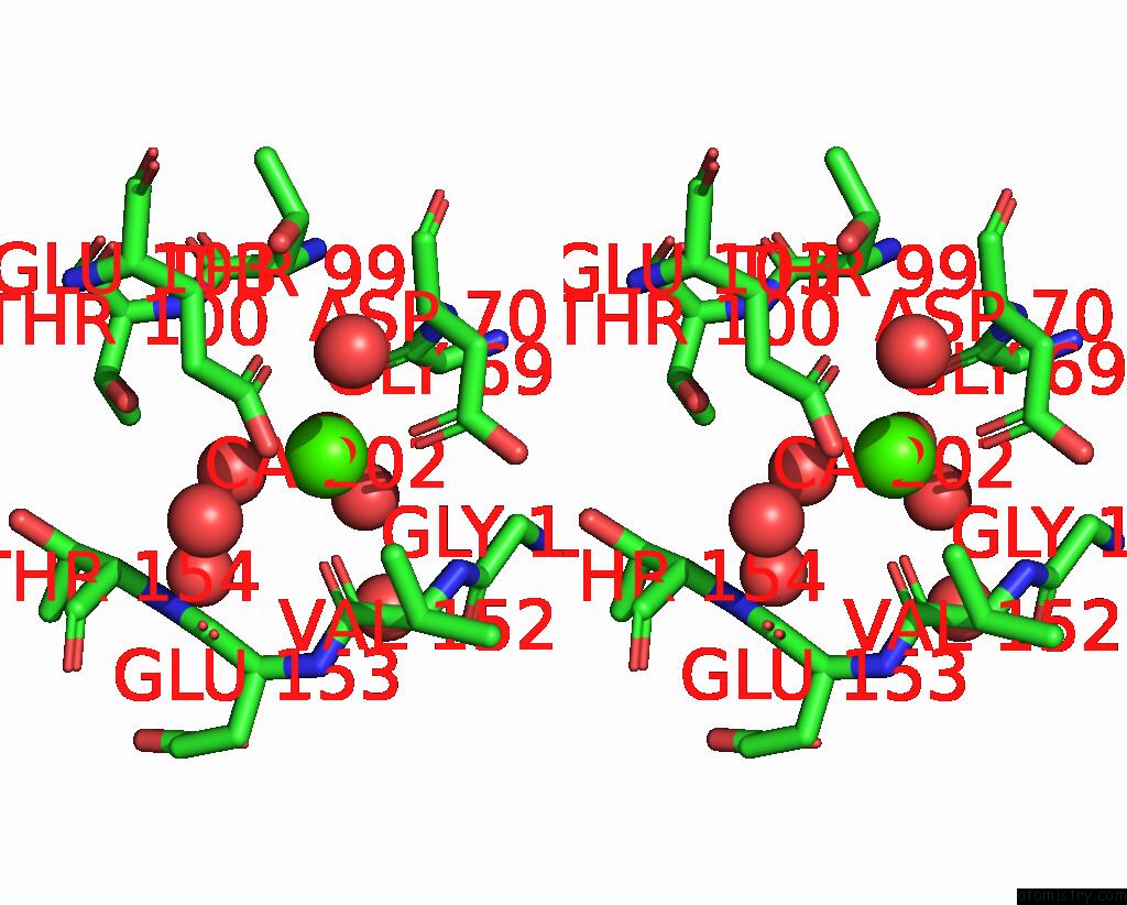
Calcium binding site 2 out of 2 in 7w4z
Go back to
Calcium binding site 2 out
of 2 in the Crystal Structure of Fragmin Domain-1 in Complex with Actin (Amppnp- Form)

Mono view
Stereo pair view
Mono view
Stereo pair view
A full contact list of Calcium with other atoms in the Ca binding
site number 2 of Crystal Structure of Fragmin Domain-1 in Complex with Actin (Amppnp- Form) within 5.0Å range:
|
Reference:
Y.Kanematsu,
A.Narita,
T.Oda,
R.Koike,
M.Ota,
Y.Takano,
K.Moritsugu,
I.Fujiwara,
K.Tanaka,
H.Komatsu,
T.Nagae,
N.Watanabe,
M.Iwasa,
Y.Maeda,
S.Takeda.
Structures and Mechanisms of Actin Atp Hydrolysis. Proc.Natl.Acad.Sci.Usa V. 119 41119 2022.
ISSN: ESSN 1091-6490
PubMed: 36252034
DOI: 10.1073/PNAS.2122641119
Page generated: Thu Jul 10 02:10:53 2025
ISSN: ESSN 1091-6490
PubMed: 36252034
DOI: 10.1073/PNAS.2122641119
Last articles
Mg in 4GMJMg in 4GNK
Mg in 4GNI
Mg in 4GN0
Mg in 4GMX
Mg in 4GME
Mg in 4GKM
Mg in 4GKR
Mg in 4GHL
Mg in 4GIU