Calcium »
PDB 7xvw-7yqs »
7ylq »
Calcium in PDB 7ylq: Crystal Structure of Microcystinase C From Sphingomonas Sp. Acm-3962 at 2.6 A Resolution
Protein crystallography data
The structure of Crystal Structure of Microcystinase C From Sphingomonas Sp. Acm-3962 at 2.6 A Resolution, PDB code: 7ylq
was solved by
X.Guo,
Z.Li,
W.Ding,
P.Yin,
L.Feng,
with X-Ray Crystallography technique. A brief refinement statistics is given in the table below:
| Resolution Low / High (Å) | 48.83 / 2.68 |
| Space group | P 61 2 2 |
| Cell size a, b, c (Å), α, β, γ (°) | 138.43, 138.43, 275.572, 90, 90, 120 |
| R / Rfree (%) | 23.1 / 26.4 |
Other elements in 7ylq:
The structure of Crystal Structure of Microcystinase C From Sphingomonas Sp. Acm-3962 at 2.6 A Resolution also contains other interesting chemical elements:
| Zinc | (Zn) | 3 atoms |
Calcium Binding Sites:
The binding sites of Calcium atom in the Crystal Structure of Microcystinase C From Sphingomonas Sp. Acm-3962 at 2.6 A Resolution
(pdb code 7ylq). This binding sites where shown within
5.0 Angstroms radius around Calcium atom.
In total only one binding site of Calcium was determined in the Crystal Structure of Microcystinase C From Sphingomonas Sp. Acm-3962 at 2.6 A Resolution, PDB code: 7ylq:
In total only one binding site of Calcium was determined in the Crystal Structure of Microcystinase C From Sphingomonas Sp. Acm-3962 at 2.6 A Resolution, PDB code: 7ylq:
Calcium binding site 1 out of 1 in 7ylq
Go back to
Calcium binding site 1 out
of 1 in the Crystal Structure of Microcystinase C From Sphingomonas Sp. Acm-3962 at 2.6 A Resolution
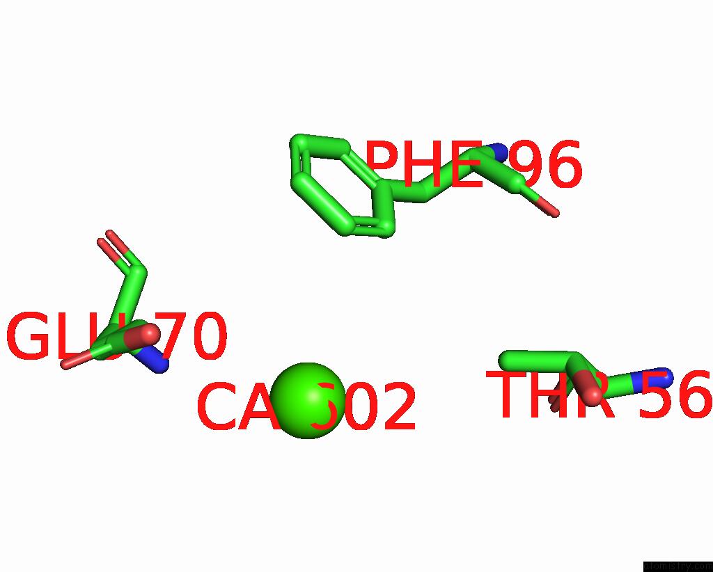
Mono view
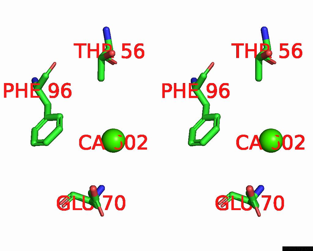
Stereo pair view
Mono view
Stereo pair view
A full contact list of Calcium with other atoms in the Ca binding
site number 1 of Crystal Structure of Microcystinase C From Sphingomonas Sp. Acm-3962 at 2.6 A Resolution within 5.0Å range:
|
Reference:
X.Guo,
Z.Li,
W.Ding,
P.Yin,
L.Feng.
Crystal Structure of Microcystin C From Sphingomonas Sp. Acm-3962 at 2.6 A Resolution To Be Published.
Page generated: Thu Jul 10 02:41:06 2025
Last articles
Mn in 9LJUMn in 9LJW
Mn in 9LJS
Mn in 9LJR
Mn in 9LJT
Mn in 9LJV
Mg in 9UA2
Mg in 9R96
Mg in 9VM1
Mg in 9P01