Calcium »
PDB 8a2u-8awz »
8a8d »
Calcium in PDB 8a8d: Atp Sulfurylase From Methanothermococcus Thermolithotrophicus - Monoclinic Form
Enzymatic activity of Atp Sulfurylase From Methanothermococcus Thermolithotrophicus - Monoclinic Form
All present enzymatic activity of Atp Sulfurylase From Methanothermococcus Thermolithotrophicus - Monoclinic Form:
2.7.7.4;
2.7.7.4;
Protein crystallography data
The structure of Atp Sulfurylase From Methanothermococcus Thermolithotrophicus - Monoclinic Form, PDB code: 8a8d
was solved by
M.Jespersen,
T.Wagner,
with X-Ray Crystallography technique. A brief refinement statistics is given in the table below:
| Resolution Low / High (Å) | 52.28 / 2.10 |
| Space group | C 1 2 1 |
| Cell size a, b, c (Å), α, β, γ (°) | 185.282, 54.525, 85.43, 90, 95.91, 90 |
| R / Rfree (%) | 19.9 / 23.9 |
Other elements in 8a8d:
The structure of Atp Sulfurylase From Methanothermococcus Thermolithotrophicus - Monoclinic Form also contains other interesting chemical elements:
| Zinc | (Zn) | 2 atoms |
Calcium Binding Sites:
The binding sites of Calcium atom in the Atp Sulfurylase From Methanothermococcus Thermolithotrophicus - Monoclinic Form
(pdb code 8a8d). This binding sites where shown within
5.0 Angstroms radius around Calcium atom.
In total 3 binding sites of Calcium where determined in the Atp Sulfurylase From Methanothermococcus Thermolithotrophicus - Monoclinic Form, PDB code: 8a8d:
Jump to Calcium binding site number: 1; 2; 3;
In total 3 binding sites of Calcium where determined in the Atp Sulfurylase From Methanothermococcus Thermolithotrophicus - Monoclinic Form, PDB code: 8a8d:
Jump to Calcium binding site number: 1; 2; 3;
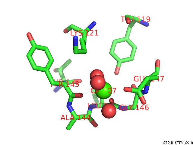
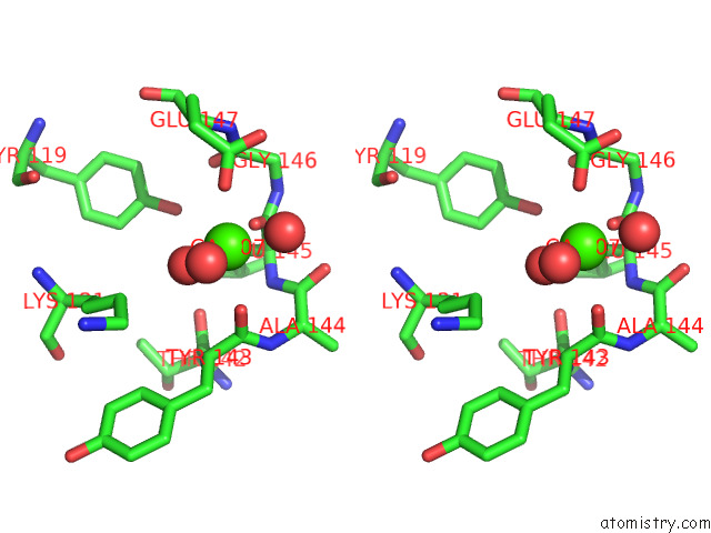
Calcium binding site 1 out of 3 in 8a8d
Go back to
Calcium binding site 1 out
of 3 in the Atp Sulfurylase From Methanothermococcus Thermolithotrophicus - Monoclinic Form
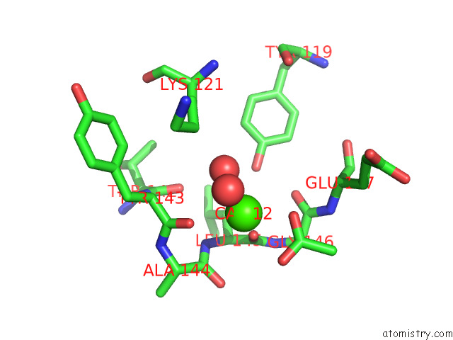
Mono view
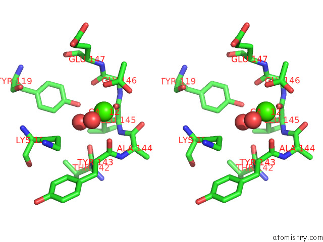
Stereo pair view
Mono view
Stereo pair view
A full contact list of Calcium with other atoms in the Ca binding
site number 1 of Atp Sulfurylase From Methanothermococcus Thermolithotrophicus - Monoclinic Form within 5.0Å range:
|
Calcium binding site 2 out of 3 in 8a8d
Go back to
Calcium binding site 2 out
of 3 in the Atp Sulfurylase From Methanothermococcus Thermolithotrophicus - Monoclinic Form
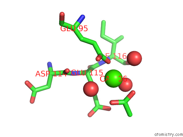
Mono view
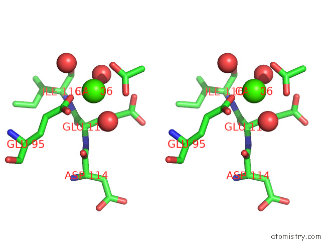
Stereo pair view
Mono view
Stereo pair view
A full contact list of Calcium with other atoms in the Ca binding
site number 2 of Atp Sulfurylase From Methanothermococcus Thermolithotrophicus - Monoclinic Form within 5.0Å range:
|
Calcium binding site 3 out of 3 in 8a8d
Go back to
Calcium binding site 3 out
of 3 in the Atp Sulfurylase From Methanothermococcus Thermolithotrophicus - Monoclinic Form
Mono view
Stereo pair view
Mono view
Stereo pair view
A full contact list of Calcium with other atoms in the Ca binding
site number 3 of Atp Sulfurylase From Methanothermococcus Thermolithotrophicus - Monoclinic Form within 5.0Å range:
|
Reference:
M.Jespersen,
T.Wagner.
How A Methanogen Assimilates Sulfate: Structural and Functional Elucidation of the Complete Sulfate-Reduction Pathway. To Be Published.
Page generated: Thu Jul 10 03:05:44 2025
Last articles
Mg in 4ZK4Mg in 4ZIR
Mg in 4ZIY
Mg in 4ZIB
Mg in 4ZI7
Mg in 4ZIL
Mg in 4ZI5
Mg in 4ZI3
Mg in 4ZIA
Mg in 4ZHQ