Calcium »
PDB 8a2u-8awz »
8at8 »
Calcium in PDB 8at8: Structure of Coproporphyrin III-Lmcpfc
Enzymatic activity of Structure of Coproporphyrin III-Lmcpfc
All present enzymatic activity of Structure of Coproporphyrin III-Lmcpfc:
4.99.1.1;
4.99.1.1;
Protein crystallography data
The structure of Structure of Coproporphyrin III-Lmcpfc, PDB code: 8at8
was solved by
T.Gabler,
S.Hofbauer,
with X-Ray Crystallography technique. A brief refinement statistics is given in the table below:
| Resolution Low / High (Å) | 36.78 / 1.51 |
| Space group | P 1 21 1 |
| Cell size a, b, c (Å), α, β, γ (°) | 37.632, 68.069, 63.036, 90, 102.22, 90 |
| R / Rfree (%) | 14.6 / 18.2 |
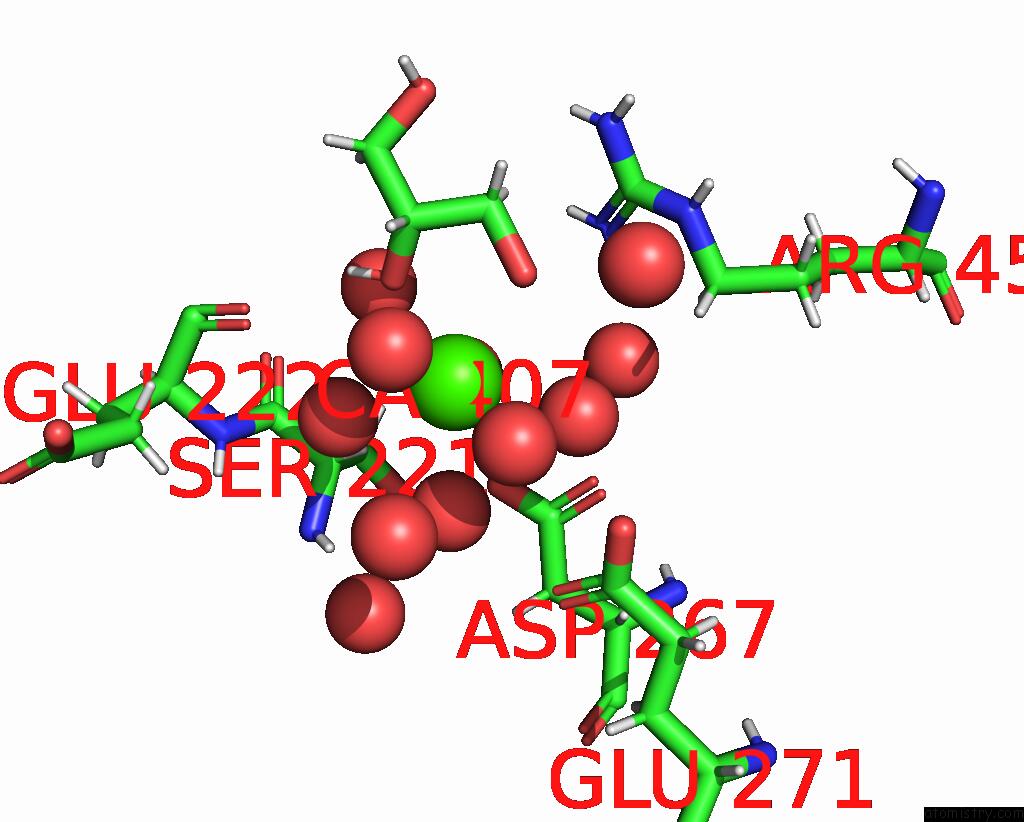
Calcium Binding Sites:
The binding sites of Calcium atom in the Structure of Coproporphyrin III-Lmcpfc
(pdb code 8at8). This binding sites where shown within
5.0 Angstroms radius around Calcium atom.
In total only one binding site of Calcium was determined in the Structure of Coproporphyrin III-Lmcpfc, PDB code: 8at8:
In total only one binding site of Calcium was determined in the Structure of Coproporphyrin III-Lmcpfc, PDB code: 8at8:
Calcium binding site 1 out of 1 in 8at8
Go back to
Calcium binding site 1 out
of 1 in the Structure of Coproporphyrin III-Lmcpfc
Mono view

Stereo pair view
Mono view
Stereo pair view
A full contact list of Calcium with other atoms in the Ca binding
site number 1 of Structure of Coproporphyrin III-Lmcpfc within 5.0Å range:
|
Reference:
A.Dali,
T.Gabler,
F.Sebastiani,
A.Destinger,
P.G.Furtmuller,
V.Pfanzagl,
M.Becucci,
G.Smulevich,
S.Hofbauer.
Active Site Architecture of Coproporphyrin Ferrochelatase with Its Physiological Substrate Coproporphyrin III: Propionate Interactions and Porphyrin Core Deformation. Protein Sci. E4534 2022.
ISSN: ESSN 1469-896X
PubMed: 36479958
DOI: 10.1002/PRO.4534
Page generated: Thu Jul 10 03:15:02 2025
ISSN: ESSN 1469-896X
PubMed: 36479958
DOI: 10.1002/PRO.4534
Last articles
Mg in 4JI5Mg in 4JI1
Mg in 4JI0
Mg in 4JI2
Mg in 4JI3
Mg in 4JHD
Mg in 4JH6
Mg in 4JH8
Mg in 4JH7
Mg in 4JH3