Calcium »
PDB 8fxt-8gpp »
8fxt »
Calcium in PDB 8fxt: Escherichia Coli Periplasmic Glucose-Binding Protein Glucose Complex: Acrylodan Conjugate Attached at W183C
Protein crystallography data
The structure of Escherichia Coli Periplasmic Glucose-Binding Protein Glucose Complex: Acrylodan Conjugate Attached at W183C, PDB code: 8fxt
was solved by
M.J.Allert,
S.Kumar,
Y.Wang,
L.S.Beese,
H.W.Hellinga,
with X-Ray Crystallography technique. A brief refinement statistics is given in the table below:
| Resolution Low / High (Å) | 24.75 / 1.53 |
| Space group | C 1 2 1 |
| Cell size a, b, c (Å), α, β, γ (°) | 119.587, 36.533, 79.905, 90, 124.13, 90 |
| R / Rfree (%) | 15.6 / 18.3 |
Calcium Binding Sites:
The binding sites of Calcium atom in the Escherichia Coli Periplasmic Glucose-Binding Protein Glucose Complex: Acrylodan Conjugate Attached at W183C
(pdb code 8fxt). This binding sites where shown within
5.0 Angstroms radius around Calcium atom.
In total only one binding site of Calcium was determined in the Escherichia Coli Periplasmic Glucose-Binding Protein Glucose Complex: Acrylodan Conjugate Attached at W183C, PDB code: 8fxt:
In total only one binding site of Calcium was determined in the Escherichia Coli Periplasmic Glucose-Binding Protein Glucose Complex: Acrylodan Conjugate Attached at W183C, PDB code: 8fxt:
Calcium binding site 1 out of 1 in 8fxt
Go back to
Calcium binding site 1 out
of 1 in the Escherichia Coli Periplasmic Glucose-Binding Protein Glucose Complex: Acrylodan Conjugate Attached at W183C
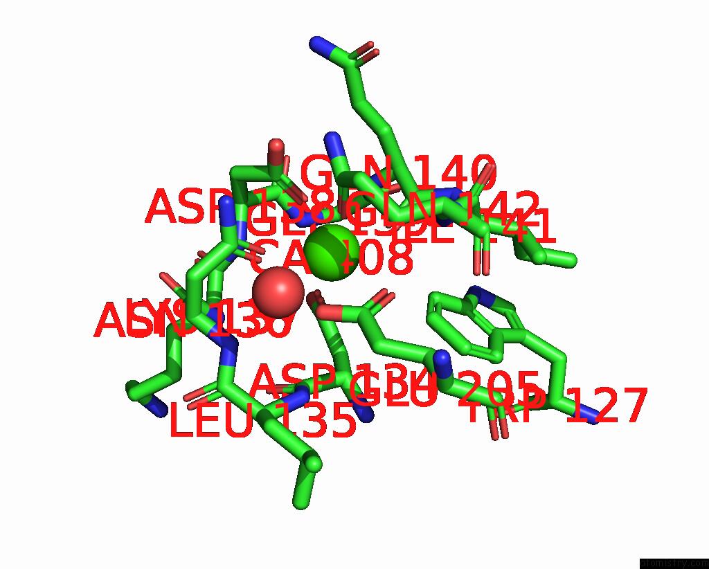
Mono view
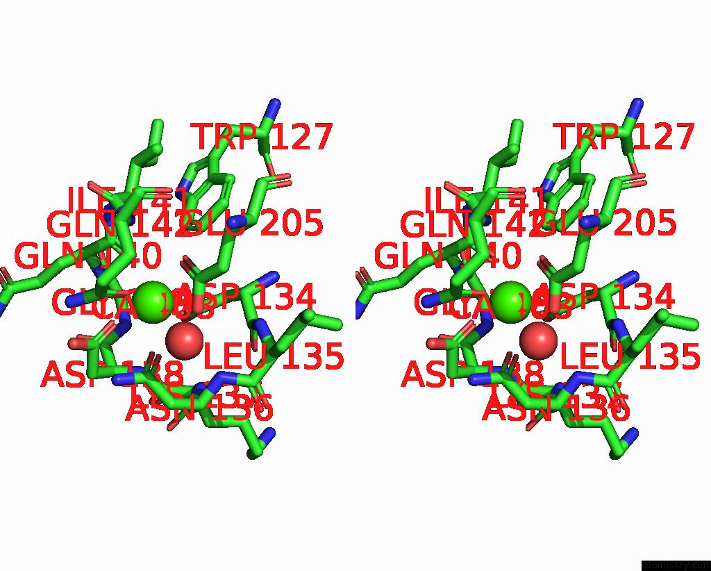
Stereo pair view
Mono view
Stereo pair view
A full contact list of Calcium with other atoms in the Ca binding
site number 1 of Escherichia Coli Periplasmic Glucose-Binding Protein Glucose Complex: Acrylodan Conjugate Attached at W183C within 5.0Å range:
|
Reference:
M.J.Allert,
S.Kumar,
Y.Wang,
L.S.Beese,
H.W.Hellinga.
Chromophore Carbonyl Twisting in Fluorescent Biosensors Encodes Direct Readout of Protein Conformations with Multicolor Switching. Commun Chem V. 6 168 2023.
ISSN: ESSN 2399-3669
PubMed: 37598249
DOI: 10.1038/S42004-023-00982-7
Page generated: Thu Jul 10 04:47:23 2025
ISSN: ESSN 2399-3669
PubMed: 37598249
DOI: 10.1038/S42004-023-00982-7
Last articles
I in 6DPEI in 6DPF
I in 6DPG
I in 6DPD
I in 6DPC
I in 6DPB
I in 6DPA
I in 6DP9
I in 6DP6
I in 6DP5