Calcium »
PDB 8gq9-8hb5 »
8h7p »
Calcium in PDB 8h7p: Crystal Structure of Aqualigase Bound with Suc-Aapf
Enzymatic activity of Crystal Structure of Aqualigase Bound with Suc-Aapf
All present enzymatic activity of Crystal Structure of Aqualigase Bound with Suc-Aapf:
3.4.21.62;
3.4.21.62;
Protein crystallography data
The structure of Crystal Structure of Aqualigase Bound with Suc-Aapf, PDB code: 8h7p
was solved by
H.Li,
M.Z.Ma,
L.J.Zhang,
L.Dai,
C.-C.Chen,
R.-T.Guo,
with X-Ray Crystallography technique. A brief refinement statistics is given in the table below:
| Resolution Low / High (Å) | 37.06 / 1.82 |
| Space group | P 1 21 1 |
| Cell size a, b, c (Å), α, β, γ (°) | 36.578, 71.68, 40.804, 90, 114.74, 90 |
| R / Rfree (%) | 13.5 / 18.9 |
Calcium Binding Sites:
The binding sites of Calcium atom in the Crystal Structure of Aqualigase Bound with Suc-Aapf
(pdb code 8h7p). This binding sites where shown within
5.0 Angstroms radius around Calcium atom.
In total only one binding site of Calcium was determined in the Crystal Structure of Aqualigase Bound with Suc-Aapf, PDB code: 8h7p:
In total only one binding site of Calcium was determined in the Crystal Structure of Aqualigase Bound with Suc-Aapf, PDB code: 8h7p:
Calcium binding site 1 out of 1 in 8h7p
Go back to
Calcium binding site 1 out
of 1 in the Crystal Structure of Aqualigase Bound with Suc-Aapf
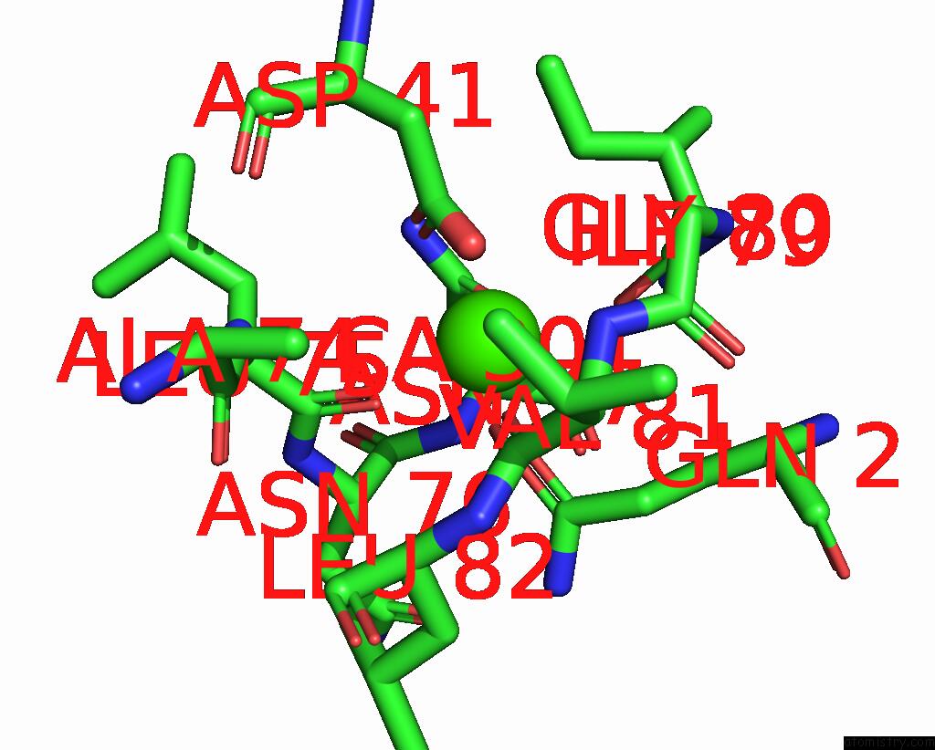
Mono view
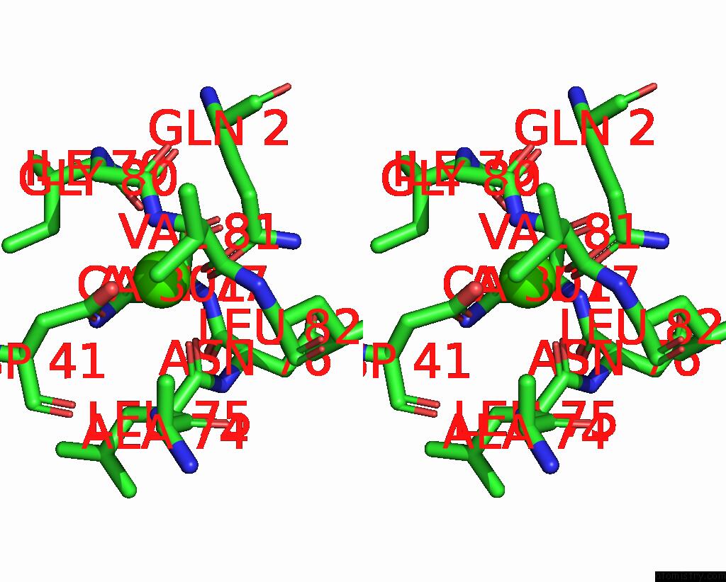
Stereo pair view
Mono view
Stereo pair view
A full contact list of Calcium with other atoms in the Ca binding
site number 1 of Crystal Structure of Aqualigase Bound with Suc-Aapf within 5.0Å range:
|
Reference:
H.Li,
M.Z.Ma,
L.J.Zhang,
L.Dai,
C.-C.Chen,
R.-T.Guo.
Crystal Structure of Aqualigase Bound with Suc-Aapf To Be Published.
Page generated: Thu Jul 10 05:01:46 2025
Last articles
Mg in 7S67Mg in 7S65
Mg in 7S66
Mg in 7S61
Mg in 7S60
Mg in 7S5Z
Mg in 7S5Y
Mg in 7S5X
Mg in 7S4X
Mg in 7RY4