Calcium »
PDB 8vh8-8wbs »
8wbs »
Calcium in PDB 8wbs: Crystal Structure of Cis-Epoxysuccinate Hydrolases Klcesh[L]-D48N Complexed with Sulfate Ions
Protein crystallography data
The structure of Crystal Structure of Cis-Epoxysuccinate Hydrolases Klcesh[L]-D48N Complexed with Sulfate Ions, PDB code: 8wbs
was solved by
S.Dong,
J.S.Xuan,
Y.G.Feng,
Q.Cui,
with X-Ray Crystallography technique. A brief refinement statistics is given in the table below:
| Resolution Low / High (Å) | 76.60 / 2.03 |
| Space group | P 21 21 2 |
| Cell size a, b, c (Å), α, β, γ (°) | 119.846, 94.426, 99.597, 90, 90, 90 |
| R / Rfree (%) | 17.8 / 20.9 |
Calcium Binding Sites:
The binding sites of Calcium atom in the Crystal Structure of Cis-Epoxysuccinate Hydrolases Klcesh[L]-D48N Complexed with Sulfate Ions
(pdb code 8wbs). This binding sites where shown within
5.0 Angstroms radius around Calcium atom.
In total 2 binding sites of Calcium where determined in the Crystal Structure of Cis-Epoxysuccinate Hydrolases Klcesh[L]-D48N Complexed with Sulfate Ions, PDB code: 8wbs:
Jump to Calcium binding site number: 1; 2;
In total 2 binding sites of Calcium where determined in the Crystal Structure of Cis-Epoxysuccinate Hydrolases Klcesh[L]-D48N Complexed with Sulfate Ions, PDB code: 8wbs:
Jump to Calcium binding site number: 1; 2;
Calcium binding site 1 out of 2 in 8wbs
Go back to
Calcium binding site 1 out
of 2 in the Crystal Structure of Cis-Epoxysuccinate Hydrolases Klcesh[L]-D48N Complexed with Sulfate Ions
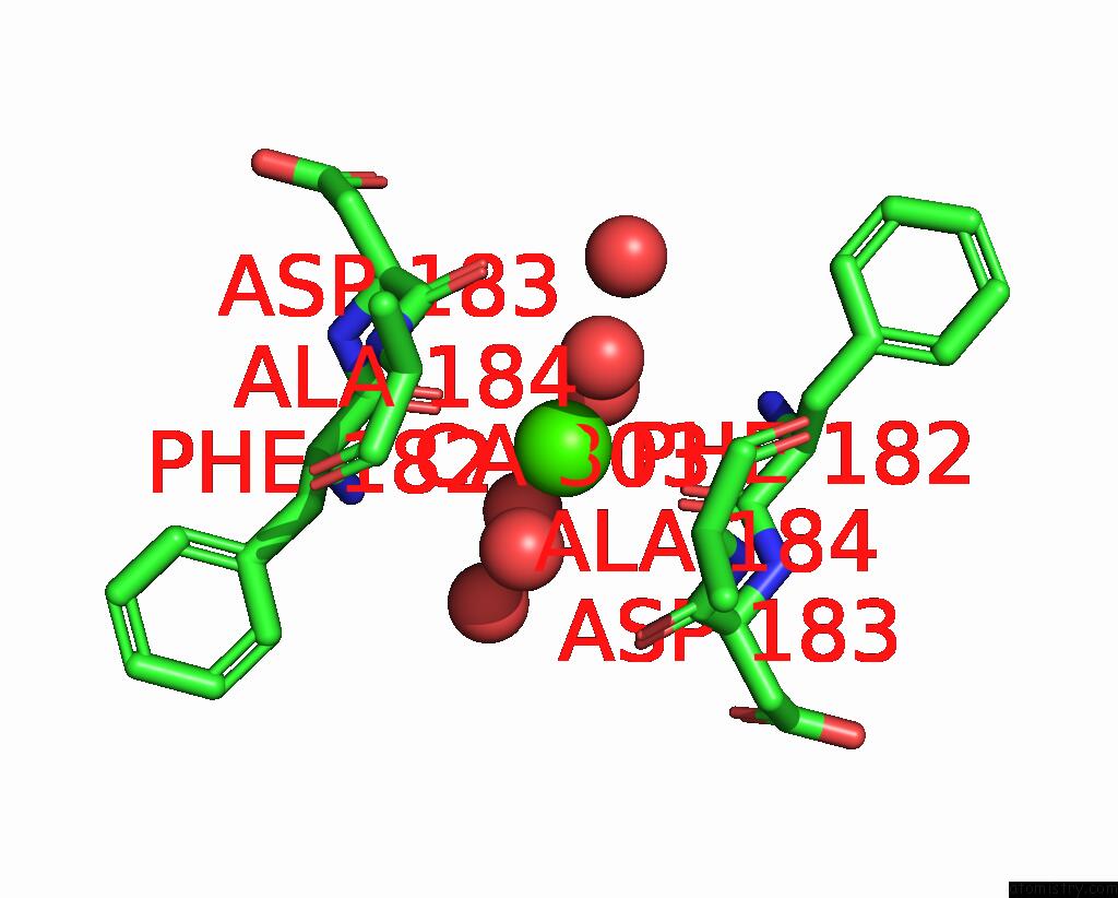
Mono view
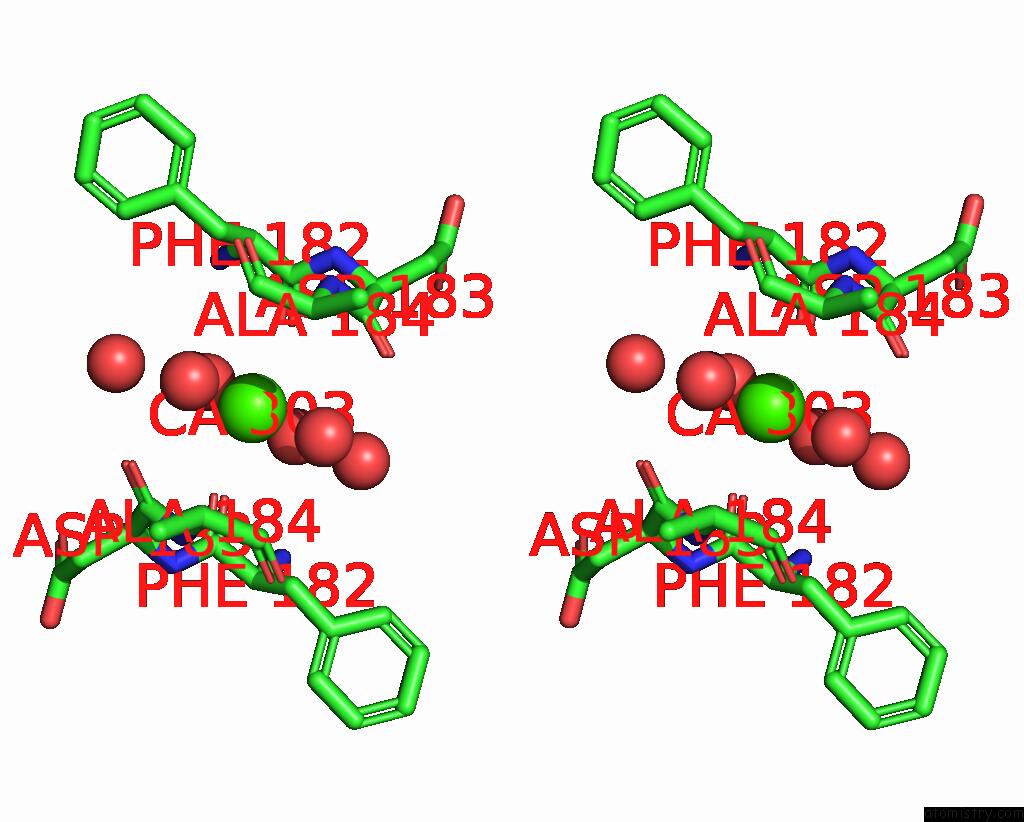
Stereo pair view
Mono view
Stereo pair view
A full contact list of Calcium with other atoms in the Ca binding
site number 1 of Crystal Structure of Cis-Epoxysuccinate Hydrolases Klcesh[L]-D48N Complexed with Sulfate Ions within 5.0Å range:
|
Calcium binding site 2 out of 2 in 8wbs
Go back to
Calcium binding site 2 out
of 2 in the Crystal Structure of Cis-Epoxysuccinate Hydrolases Klcesh[L]-D48N Complexed with Sulfate Ions
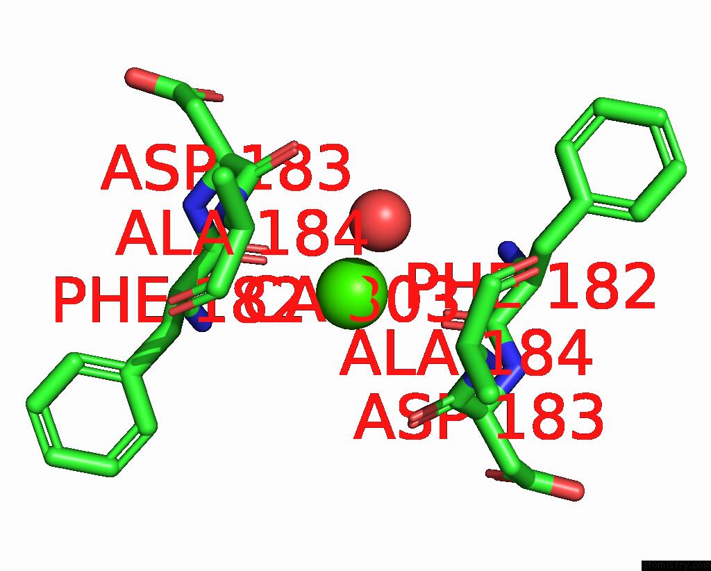
Mono view
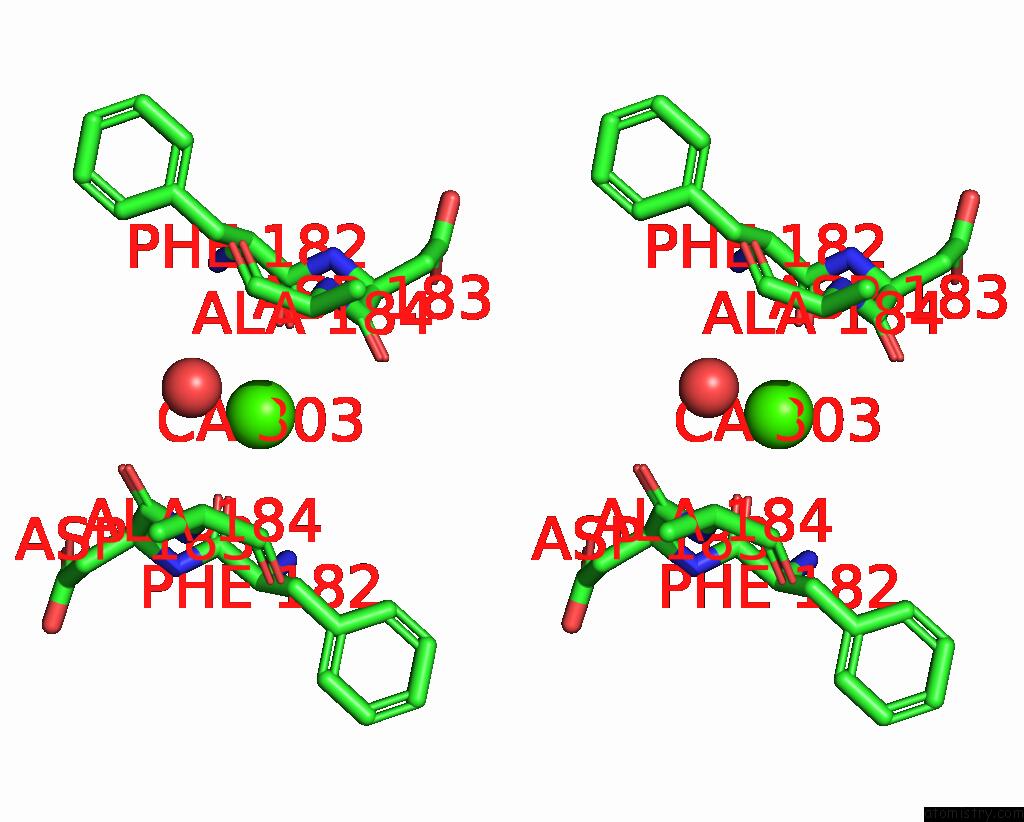
Stereo pair view
Mono view
Stereo pair view
A full contact list of Calcium with other atoms in the Ca binding
site number 2 of Crystal Structure of Cis-Epoxysuccinate Hydrolases Klcesh[L]-D48N Complexed with Sulfate Ions within 5.0Å range:
|
Reference:
S.Dong,
J.Xuan,
Y.Feng,
Q.Cui.
Deciphering the Stereo-Specific Catalytic Mechanisms of Cis-Epoxysuccinate Hydrolases Producing L(+)-Tartaric Acid. J.Biol.Chem. 05635 2024.
ISSN: ESSN 1083-351X
PubMed: 38199576
DOI: 10.1016/J.JBC.2024.105635
Page generated: Thu Jul 10 08:07:45 2025
ISSN: ESSN 1083-351X
PubMed: 38199576
DOI: 10.1016/J.JBC.2024.105635
Last articles
Mn in 9LJUMn in 9LJW
Mn in 9LJS
Mn in 9LJR
Mn in 9LJT
Mn in 9LJV
Mg in 9UA2
Mg in 9R96
Mg in 9VM1
Mg in 9P01