Calcium »
PDB 8wpl-8xvr »
8xvp »
Calcium in PDB 8xvp: Crystal Structure of Inulosucrase From Lactobacillus Reuteri 121
Enzymatic activity of Crystal Structure of Inulosucrase From Lactobacillus Reuteri 121
All present enzymatic activity of Crystal Structure of Inulosucrase From Lactobacillus Reuteri 121:
2.4.1.9;
2.4.1.9;
Protein crystallography data
The structure of Crystal Structure of Inulosucrase From Lactobacillus Reuteri 121, PDB code: 8xvp
was solved by
D.Ni,
X.Hou,
M.Cheng,
W.Xu,
Y.Rao,
W.Mu,
with X-Ray Crystallography technique. A brief refinement statistics is given in the table below:
| Resolution Low / High (Å) | 49.42 / 1.98 |
| Space group | P 21 21 21 |
| Cell size a, b, c (Å), α, β, γ (°) | 69.91, 98.69, 139.49, 90, 90, 90 |
| R / Rfree (%) | 20 / 24.5 |
Calcium Binding Sites:
The binding sites of Calcium atom in the Crystal Structure of Inulosucrase From Lactobacillus Reuteri 121
(pdb code 8xvp). This binding sites where shown within
5.0 Angstroms radius around Calcium atom.
In total 6 binding sites of Calcium where determined in the Crystal Structure of Inulosucrase From Lactobacillus Reuteri 121, PDB code: 8xvp:
Jump to Calcium binding site number: 1; 2; 3; 4; 5; 6;
In total 6 binding sites of Calcium where determined in the Crystal Structure of Inulosucrase From Lactobacillus Reuteri 121, PDB code: 8xvp:
Jump to Calcium binding site number: 1; 2; 3; 4; 5; 6;
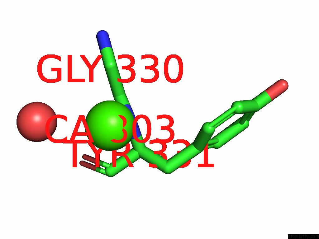
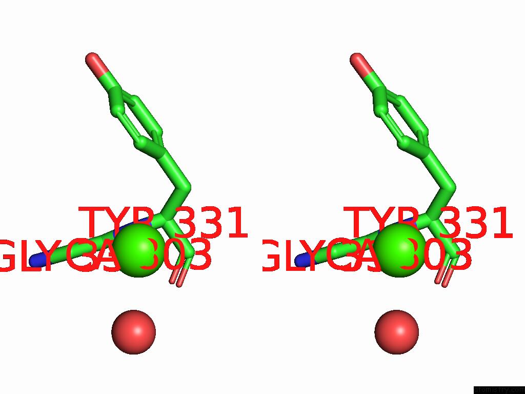
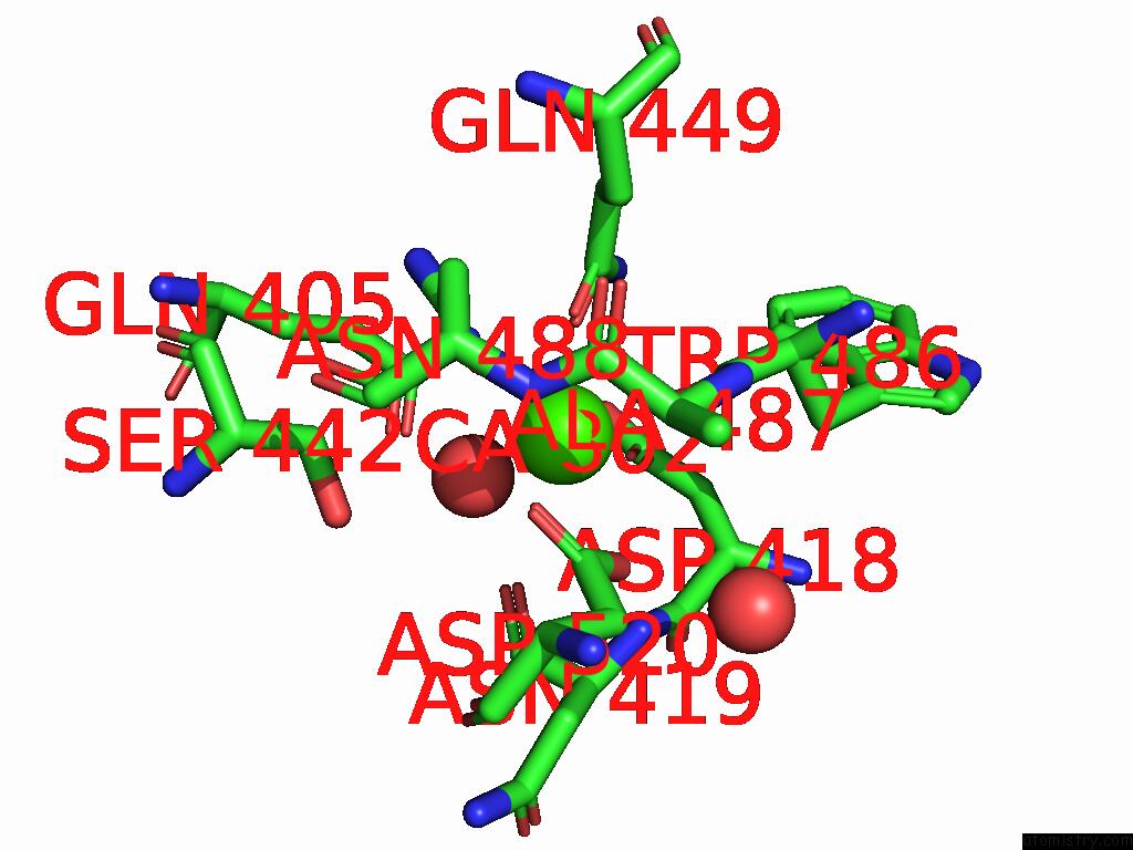
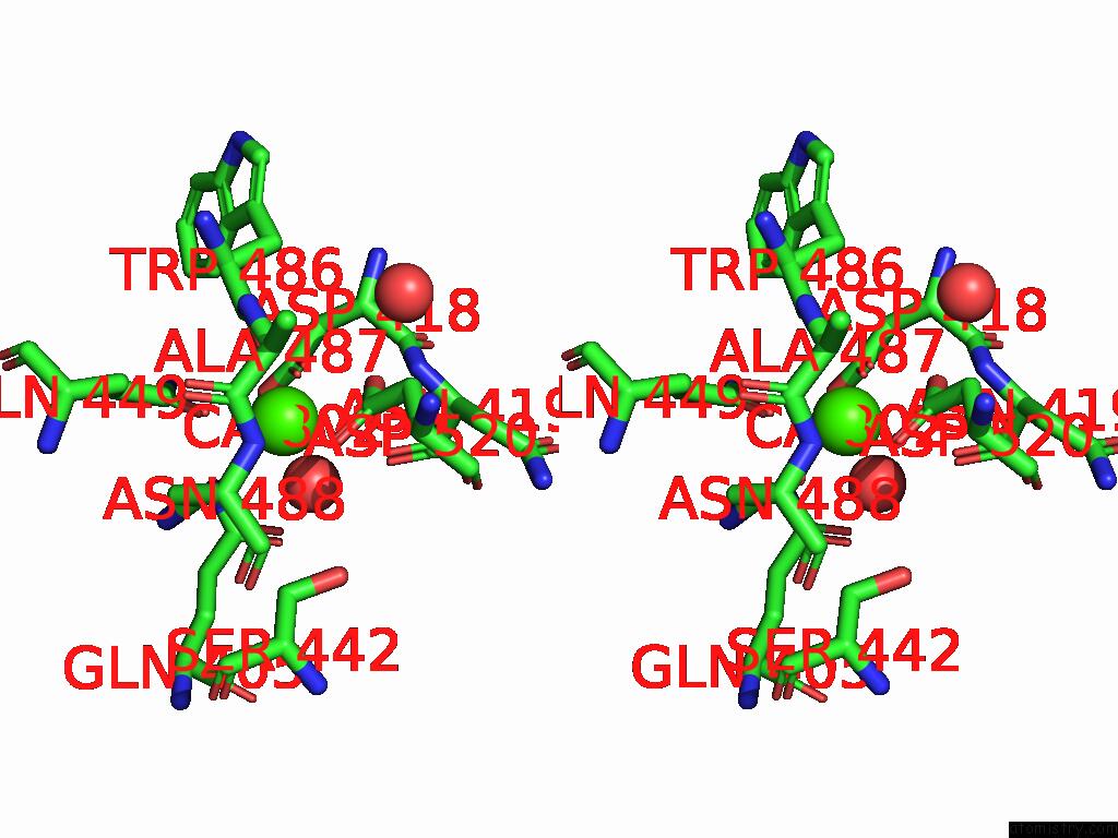
Calcium binding site 1 out of 6 in 8xvp
Go back to
Calcium binding site 1 out
of 6 in the Crystal Structure of Inulosucrase From Lactobacillus Reuteri 121
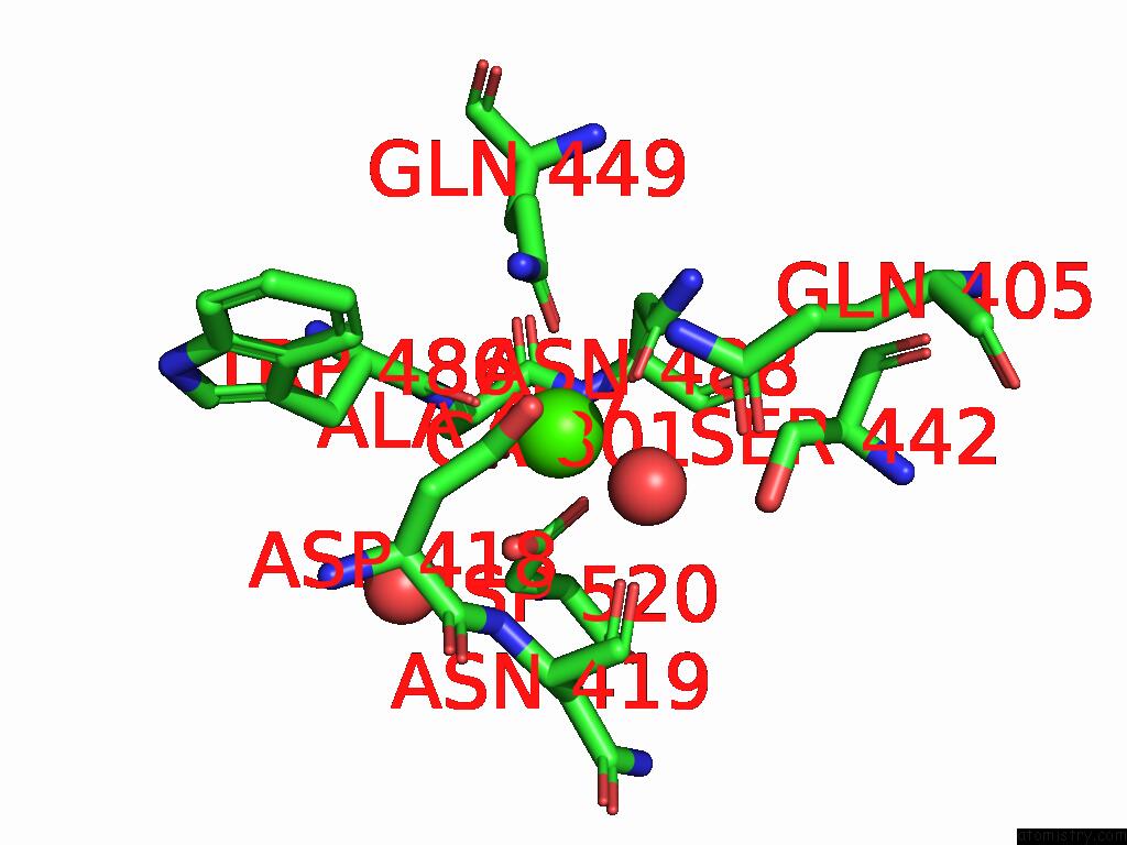
Mono view
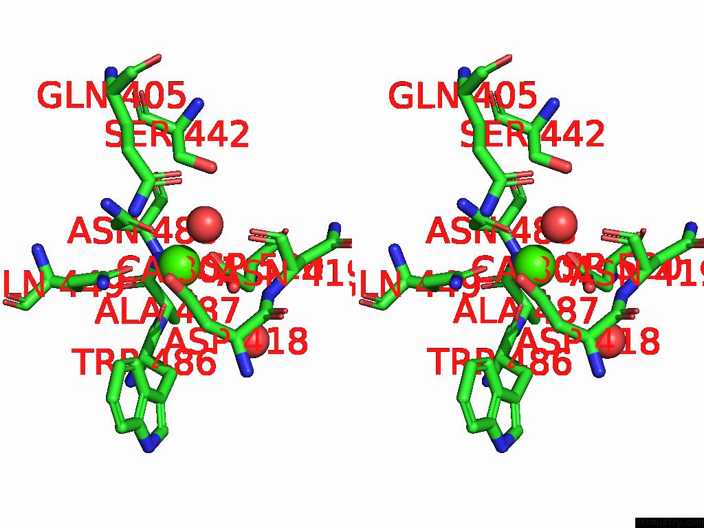
Stereo pair view
Mono view
Stereo pair view
A full contact list of Calcium with other atoms in the Ca binding
site number 1 of Crystal Structure of Inulosucrase From Lactobacillus Reuteri 121 within 5.0Å range:
|
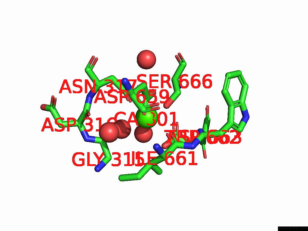
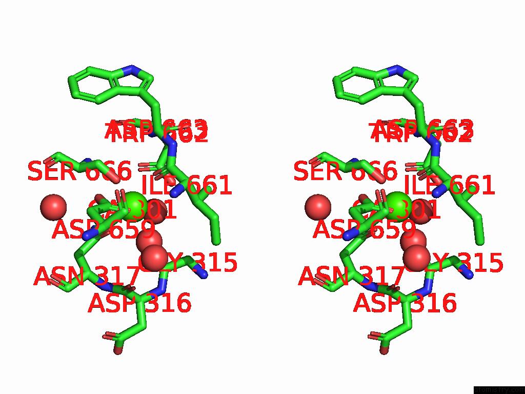
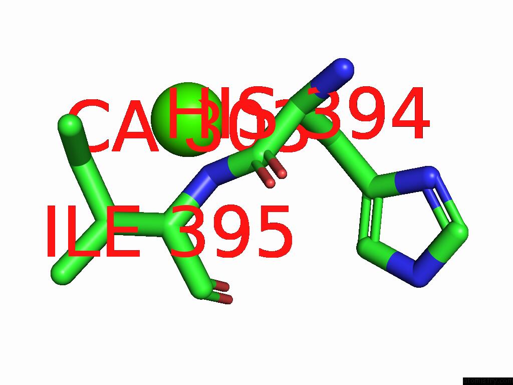
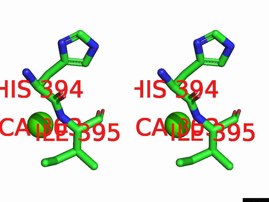
Calcium binding site 2 out of 6 in 8xvp
Go back to
Calcium binding site 2 out
of 6 in the Crystal Structure of Inulosucrase From Lactobacillus Reuteri 121
Mono view
Stereo pair view
Mono view
Stereo pair view
A full contact list of Calcium with other atoms in the Ca binding
site number 2 of Crystal Structure of Inulosucrase From Lactobacillus Reuteri 121 within 5.0Å range:
|
Calcium binding site 3 out of 6 in 8xvp
Go back to
Calcium binding site 3 out
of 6 in the Crystal Structure of Inulosucrase From Lactobacillus Reuteri 121
Mono view
Stereo pair view
Mono view
Stereo pair view
A full contact list of Calcium with other atoms in the Ca binding
site number 3 of Crystal Structure of Inulosucrase From Lactobacillus Reuteri 121 within 5.0Å range:
|
Calcium binding site 4 out of 6 in 8xvp
Go back to
Calcium binding site 4 out
of 6 in the Crystal Structure of Inulosucrase From Lactobacillus Reuteri 121
Mono view
Stereo pair view
Mono view
Stereo pair view
A full contact list of Calcium with other atoms in the Ca binding
site number 4 of Crystal Structure of Inulosucrase From Lactobacillus Reuteri 121 within 5.0Å range:
|
Calcium binding site 5 out of 6 in 8xvp
Go back to
Calcium binding site 5 out
of 6 in the Crystal Structure of Inulosucrase From Lactobacillus Reuteri 121
Mono view
Stereo pair view
Mono view
Stereo pair view
A full contact list of Calcium with other atoms in the Ca binding
site number 5 of Crystal Structure of Inulosucrase From Lactobacillus Reuteri 121 within 5.0Å range:
|
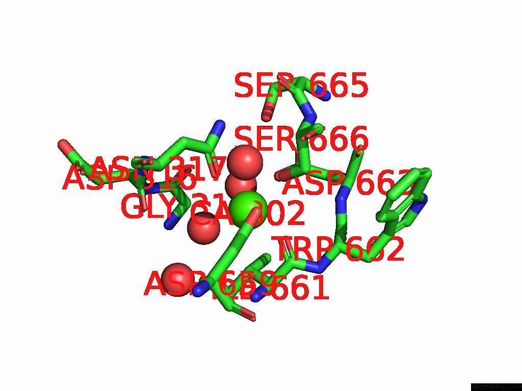
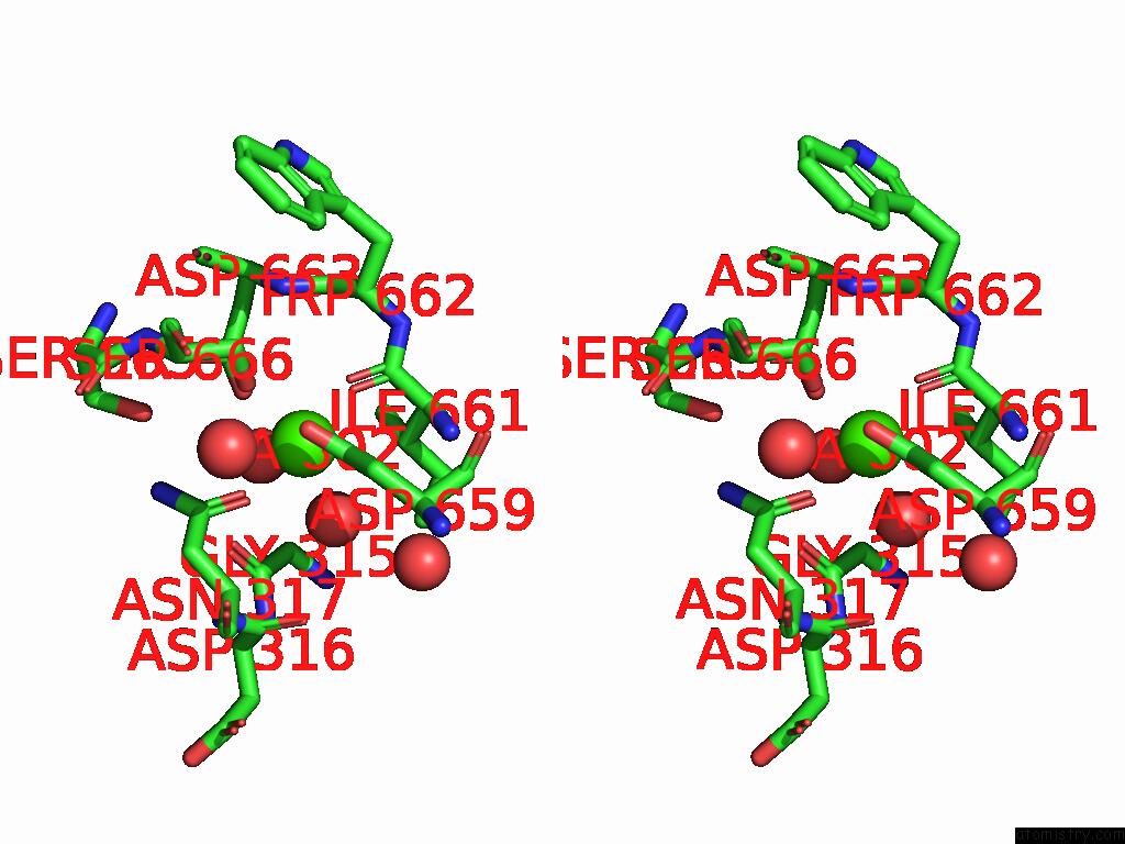
Calcium binding site 6 out of 6 in 8xvp
Go back to
Calcium binding site 6 out
of 6 in the Crystal Structure of Inulosucrase From Lactobacillus Reuteri 121
Mono view
Stereo pair view
Mono view
Stereo pair view
A full contact list of Calcium with other atoms in the Ca binding
site number 6 of Crystal Structure of Inulosucrase From Lactobacillus Reuteri 121 within 5.0Å range:
|
Reference:
D.Ni,
X.Hou,
M.Cheng,
W.Xu,
Y.Rao,
W.Mu.
Structure-Guided Tunnel Engineering to Reveal the Molecular Basis of Sugar Chain Extension of Inulosucrase To Be Published.
Page generated: Sat Feb 8 16:11:43 2025
Last articles
Zn in 9MJ5Zn in 9HNW
Zn in 9G0L
Zn in 9FNE
Zn in 9DZN
Zn in 9E0I
Zn in 9D32
Zn in 9DAK
Zn in 8ZXC
Zn in 8ZUF