Calcium »
PDB 8zlw-9awo »
9avz »
Calcium in PDB 9avz: Crystal Structure of Trypsin at 100 Kelvin with Benzamidine (Triplicate)
Enzymatic activity of Crystal Structure of Trypsin at 100 Kelvin with Benzamidine (Triplicate)
All present enzymatic activity of Crystal Structure of Trypsin at 100 Kelvin with Benzamidine (Triplicate):
3.4.21.4;
3.4.21.4;
Protein crystallography data
The structure of Crystal Structure of Trypsin at 100 Kelvin with Benzamidine (Triplicate), PDB code: 9avz
was solved by
L.M.T.R.Lima,
F.De Sa Ribeiro,
with X-Ray Crystallography technique. A brief refinement statistics is given in the table below:
| Resolution Low / High (Å) | 24.53 / 1.50 |
| Space group | P 21 21 21 |
| Cell size a, b, c (Å), α, β, γ (°) | 54.095, 58.074, 66.573, 90, 90, 90 |
| R / Rfree (%) | 20.6 / 23.3 |
Calcium Binding Sites:
The binding sites of Calcium atom in the Crystal Structure of Trypsin at 100 Kelvin with Benzamidine (Triplicate)
(pdb code 9avz). This binding sites where shown within
5.0 Angstroms radius around Calcium atom.
In total only one binding site of Calcium was determined in the Crystal Structure of Trypsin at 100 Kelvin with Benzamidine (Triplicate), PDB code: 9avz:
In total only one binding site of Calcium was determined in the Crystal Structure of Trypsin at 100 Kelvin with Benzamidine (Triplicate), PDB code: 9avz:
Calcium binding site 1 out of 1 in 9avz
Go back to
Calcium binding site 1 out
of 1 in the Crystal Structure of Trypsin at 100 Kelvin with Benzamidine (Triplicate)
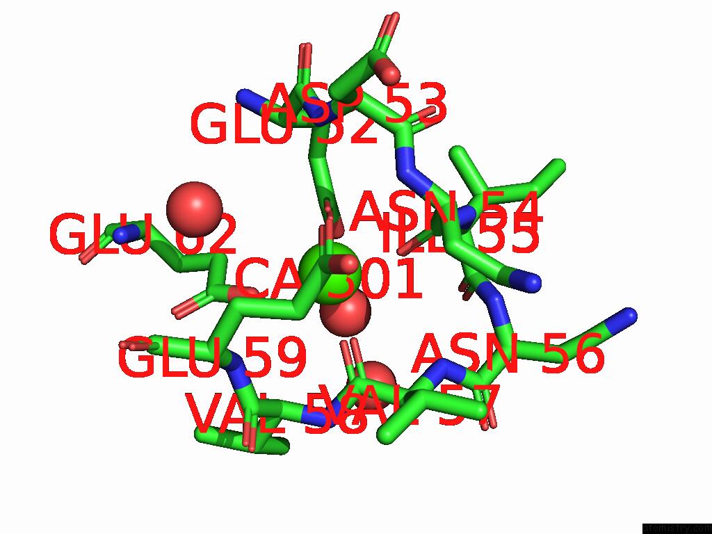
Mono view
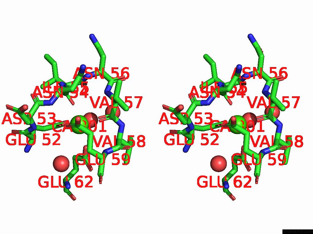
Stereo pair view
Mono view
Stereo pair view
A full contact list of Calcium with other atoms in the Ca binding
site number 1 of Crystal Structure of Trypsin at 100 Kelvin with Benzamidine (Triplicate) within 5.0Å range:
|
Reference:
L.M.T.R.Lima,
F.De Sa Ribeiro.
Crystal Structure of Trypsin at 100 Kelvin with Benzamidine (Triplicate) To Be Published.
Page generated: Thu Jul 10 08:49:40 2025
Last articles
Mg in 8EZBMg in 8EZF
Mg in 8EYE
Mg in 8EZ2
Mg in 8EXX
Mg in 8EX3
Mg in 8EXY
Mg in 8EVZ
Mg in 8EXW
Mg in 8EWZ