Calcium »
PDB 9ibz-9kzc »
9jlm »
Calcium in PDB 9jlm: Crystal Structure of Aldolase Atob 1.9A
Protein crystallography data
The structure of Crystal Structure of Aldolase Atob 1.9A, PDB code: 9jlm
was solved by
K.Ma,
A.Fan,
W.Lin,
with X-Ray Crystallography technique. A brief refinement statistics is given in the table below:
| Resolution Low / High (Å) | 41.08 / 1.90 |
| Space group | P 1 21 1 |
| Cell size a, b, c (Å), α, β, γ (°) | 51.228, 70.074, 51.834, 90, 101.95, 90 |
| R / Rfree (%) | 21 / 26.3 |
Calcium Binding Sites:
The binding sites of Calcium atom in the Crystal Structure of Aldolase Atob 1.9A
(pdb code 9jlm). This binding sites where shown within
5.0 Angstroms radius around Calcium atom.
In total 2 binding sites of Calcium where determined in the Crystal Structure of Aldolase Atob 1.9A, PDB code: 9jlm:
Jump to Calcium binding site number: 1; 2;
In total 2 binding sites of Calcium where determined in the Crystal Structure of Aldolase Atob 1.9A, PDB code: 9jlm:
Jump to Calcium binding site number: 1; 2;
Calcium binding site 1 out of 2 in 9jlm
Go back to
Calcium binding site 1 out
of 2 in the Crystal Structure of Aldolase Atob 1.9A
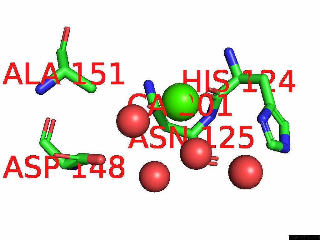
Mono view
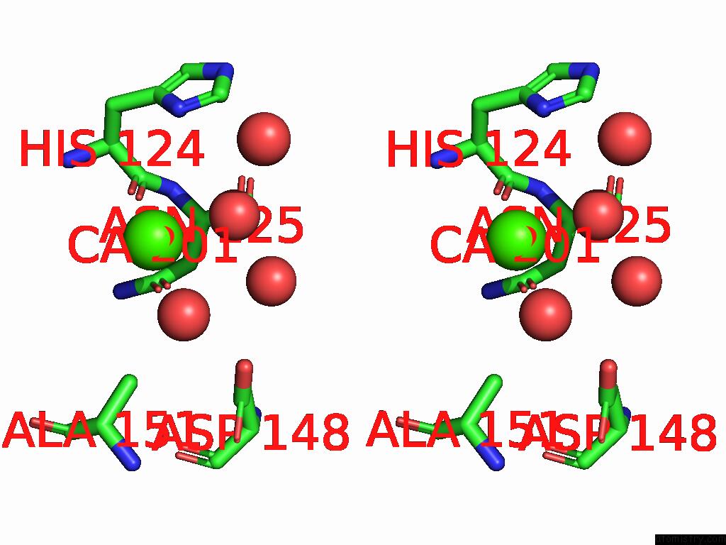
Stereo pair view
Mono view
Stereo pair view
A full contact list of Calcium with other atoms in the Ca binding
site number 1 of Crystal Structure of Aldolase Atob 1.9A within 5.0Å range:
|
Calcium binding site 2 out of 2 in 9jlm
Go back to
Calcium binding site 2 out
of 2 in the Crystal Structure of Aldolase Atob 1.9A
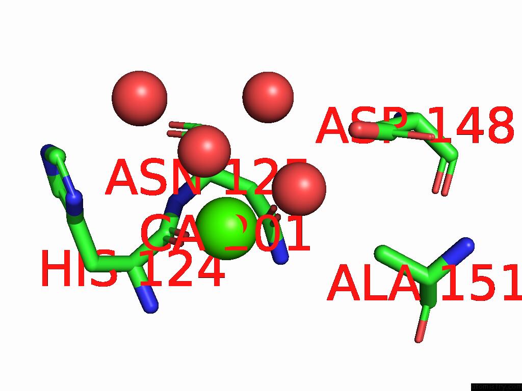
Mono view
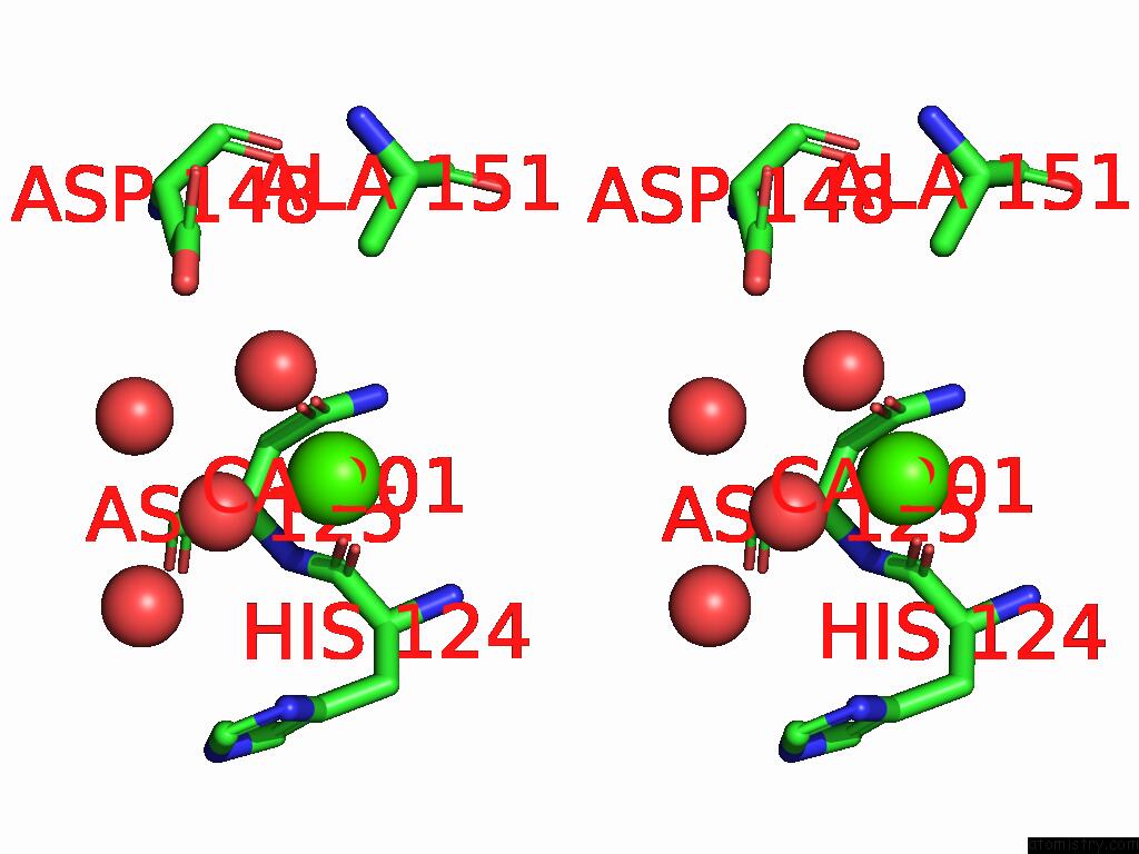
Stereo pair view
Mono view
Stereo pair view
A full contact list of Calcium with other atoms in the Ca binding
site number 2 of Crystal Structure of Aldolase Atob 1.9A within 5.0Å range:
|
Reference:
K.Ma,
J.Liu,
Z.Huang,
M.Wu,
D.Liu,
J.Ren,
A.Fan,
W.Lin.
Three-Dimensional Structural Alignment Based Discovery and Molecular Basis of Atob, Catalyzing Linear Tetracyclic Formation. Chem Sci V. 15 18490 2024.
ISSN: ISSN 2041-6520
PubMed: 39430940
DOI: 10.1039/D4SC05590J
Page generated: Thu Jul 10 09:56:28 2025
ISSN: ISSN 2041-6520
PubMed: 39430940
DOI: 10.1039/D4SC05590J
Last articles
Fe in 2YXOFe in 2YRS
Fe in 2YXC
Fe in 2YNM
Fe in 2YVJ
Fe in 2YP1
Fe in 2YU2
Fe in 2YU1
Fe in 2YQB
Fe in 2YOO