Calcium »
PDB 1spu-1t5s »
1sxn »
Calcium in PDB 1sxn: Reduced Bovine Superoxide Dismutase at pH 5.0
Enzymatic activity of Reduced Bovine Superoxide Dismutase at pH 5.0
All present enzymatic activity of Reduced Bovine Superoxide Dismutase at pH 5.0:
1.15.1.1;
1.15.1.1;
Protein crystallography data
The structure of Reduced Bovine Superoxide Dismutase at pH 5.0, PDB code: 1sxn
was solved by
M.Ferraroni,
W.R.Rypniewski,
B.Bruni,
P.Orioli,
K.S.Wilson,
S.Mangani,
with X-Ray Crystallography technique. A brief refinement statistics is given in the table below:
| Resolution Low / High (Å) | 10.00 / 1.90 |
| Space group | C 2 2 21 |
| Cell size a, b, c (Å), α, β, γ (°) | 104.690, 197.800, 50.830, 90.00, 90.00, 90.00 |
| R / Rfree (%) | 18 / n/a |
Other elements in 1sxn:
The structure of Reduced Bovine Superoxide Dismutase at pH 5.0 also contains other interesting chemical elements:
| Copper | (Cu) | 2 atoms |
| Zinc | (Zn) | 2 atoms |
Calcium Binding Sites:
The binding sites of Calcium atom in the Reduced Bovine Superoxide Dismutase at pH 5.0
(pdb code 1sxn). This binding sites where shown within
5.0 Angstroms radius around Calcium atom.
In total 2 binding sites of Calcium where determined in the Reduced Bovine Superoxide Dismutase at pH 5.0, PDB code: 1sxn:
Jump to Calcium binding site number: 1; 2;
In total 2 binding sites of Calcium where determined in the Reduced Bovine Superoxide Dismutase at pH 5.0, PDB code: 1sxn:
Jump to Calcium binding site number: 1; 2;
Calcium binding site 1 out of 2 in 1sxn
Go back to
Calcium binding site 1 out
of 2 in the Reduced Bovine Superoxide Dismutase at pH 5.0
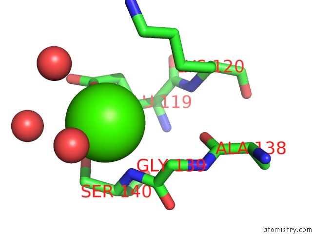
Mono view
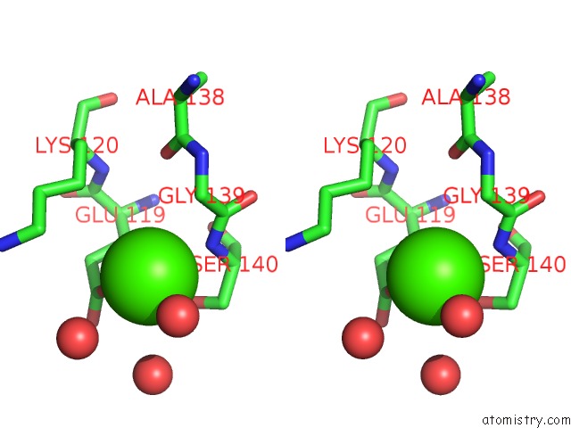
Stereo pair view
Mono view
Stereo pair view
A full contact list of Calcium with other atoms in the Ca binding
site number 1 of Reduced Bovine Superoxide Dismutase at pH 5.0 within 5.0Å range:
|
Calcium binding site 2 out of 2 in 1sxn
Go back to
Calcium binding site 2 out
of 2 in the Reduced Bovine Superoxide Dismutase at pH 5.0
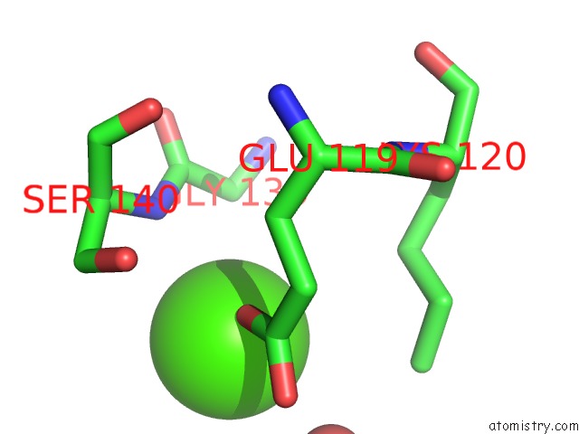
Mono view
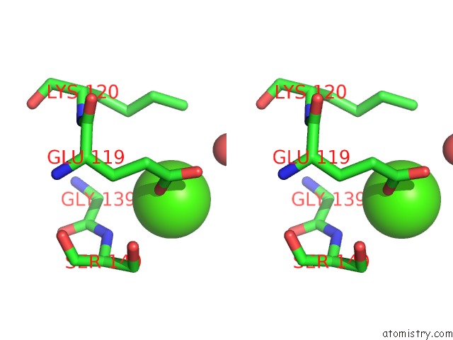
Stereo pair view
Mono view
Stereo pair view
A full contact list of Calcium with other atoms in the Ca binding
site number 2 of Reduced Bovine Superoxide Dismutase at pH 5.0 within 5.0Å range:
|
Reference:
M.Ferraroni,
W.R.Rypniewski,
B.Bruni,
P.Orioli,
S.Mangani.
Crystallographic Determination of Reduced Bovine Superoxide Dismutase at pH 5.0 and of Anion Binding to Its Active Site. J.Biol.Inorg.Chem. V. 3 411 1998.
ISSN: ISSN 0949-8257
Page generated: Tue Jul 8 02:07:37 2025
ISSN: ISSN 0949-8257
Last articles
Ca in 7K9OCa in 7K9N
Ca in 7K6C
Ca in 7K96
Ca in 7K72
Ca in 7K1R
Ca in 7K66
Ca in 7K4W
Ca in 7K4O
Ca in 7K4C