Calcium »
PDB 7k1r-7kl5 »
7k66 »
Calcium in PDB 7k66: Structure of Blood Coagulation Factor VIII in Complex with An Anti-C1 Domain Pathogenic Antibody Inhibitor
Protein crystallography data
The structure of Structure of Blood Coagulation Factor VIII in Complex with An Anti-C1 Domain Pathogenic Antibody Inhibitor, PDB code: 7k66
was solved by
K.C.Childers,
J.Gish,
L.Jarvis,
S.Peters,
C.Garrels,
I.W.Smith,
H.T.Spencer,
P.C.Spiegel,
with X-Ray Crystallography technique. A brief refinement statistics is given in the table below:
| Resolution Low / High (Å) | 49.49 / 3.92 |
| Space group | P 41 21 2 |
| Cell size a, b, c (Å), α, β, γ (°) | 116.721, 116.721, 371.057, 90.00, 90.00, 90.00 |
| R / Rfree (%) | 22.6 / 32.2 |
Other elements in 7k66:
The structure of Structure of Blood Coagulation Factor VIII in Complex with An Anti-C1 Domain Pathogenic Antibody Inhibitor also contains other interesting chemical elements:
| Copper | (Cu) | 1 atom |
| Zinc | (Zn) | 1 atom |
Calcium Binding Sites:
The binding sites of Calcium atom in the Structure of Blood Coagulation Factor VIII in Complex with An Anti-C1 Domain Pathogenic Antibody Inhibitor
(pdb code 7k66). This binding sites where shown within
5.0 Angstroms radius around Calcium atom.
In total only one binding site of Calcium was determined in the Structure of Blood Coagulation Factor VIII in Complex with An Anti-C1 Domain Pathogenic Antibody Inhibitor, PDB code: 7k66:
In total only one binding site of Calcium was determined in the Structure of Blood Coagulation Factor VIII in Complex with An Anti-C1 Domain Pathogenic Antibody Inhibitor, PDB code: 7k66:
Calcium binding site 1 out of 1 in 7k66
Go back to
Calcium binding site 1 out
of 1 in the Structure of Blood Coagulation Factor VIII in Complex with An Anti-C1 Domain Pathogenic Antibody Inhibitor
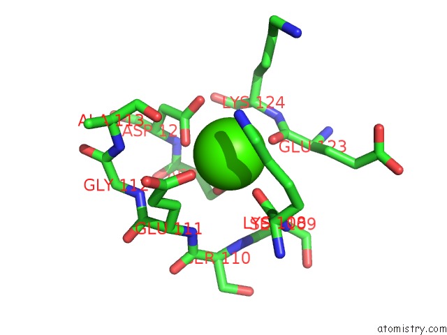
Mono view
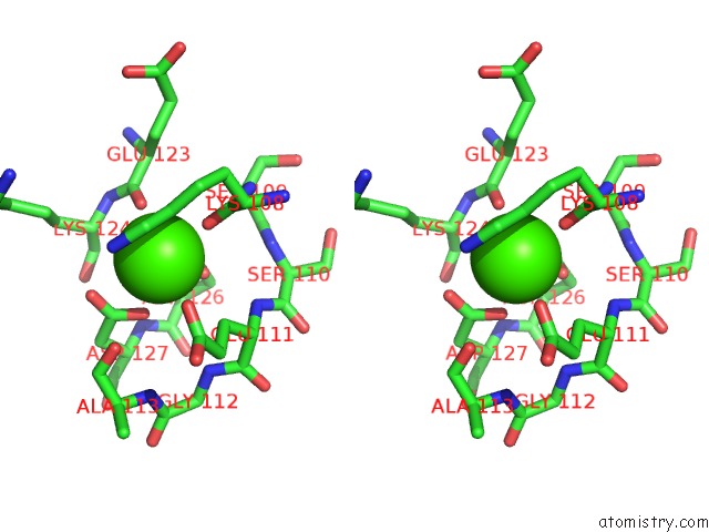
Stereo pair view
Mono view
Stereo pair view
A full contact list of Calcium with other atoms in the Ca binding
site number 1 of Structure of Blood Coagulation Factor VIII in Complex with An Anti-C1 Domain Pathogenic Antibody Inhibitor within 5.0Å range:
|
Reference:
J.Gish,
L.Jarvis,
K.C.Childers,
S.Peters,
C.Garrels,
I.W.Smith,
H.T.Spencer,
P.Lollar,
C.B.Doering,
P.C.Spiegel.
Structure of Blood Coagulation Factor VIII in Complex with An Anti-C1 Domain Pathogenic Antibody Inhibitor To Be Published.
Page generated: Wed Jul 9 22:50:39 2025
Last articles
Fe in 9ONMFe in 9NSV
Fe in 9O8U
Fe in 9N5V
Fe in 9NSE
Fe in 9NQU
Fe in 9NP6
Fe in 9NH3
Fe in 9NEA
Fe in 9NE9