Calcium »
PDB 2b04-2bib »
2bax »
Calcium in PDB 2bax: Atomic Resolution Structure of the Double Mutant (K53,56M) of Bovine Pancreatic Phospholipase A2
Enzymatic activity of Atomic Resolution Structure of the Double Mutant (K53,56M) of Bovine Pancreatic Phospholipase A2
All present enzymatic activity of Atomic Resolution Structure of the Double Mutant (K53,56M) of Bovine Pancreatic Phospholipase A2:
3.1.1.4;
3.1.1.4;
Protein crystallography data
The structure of Atomic Resolution Structure of the Double Mutant (K53,56M) of Bovine Pancreatic Phospholipase A2, PDB code: 2bax
was solved by
K.Sekar,
M.Yogavel,
D.Velmurugan,
Z.Dauter,
M.Dauter,
M.D.Tsai,
with X-Ray Crystallography technique. A brief refinement statistics is given in the table below:
| Resolution Low / High (Å) | 20.00 / 1.10 |
| Space group | P 31 2 1 |
| Cell size a, b, c (Å), α, β, γ (°) | 46.057, 46.057, 101.138, 90.00, 90.00, 120.00 |
| R / Rfree (%) | 11.9 / 15.6 |
Other elements in 2bax:
The structure of Atomic Resolution Structure of the Double Mutant (K53,56M) of Bovine Pancreatic Phospholipase A2 also contains other interesting chemical elements:
| Chlorine | (Cl) | 1 atom |
Calcium Binding Sites:
The binding sites of Calcium atom in the Atomic Resolution Structure of the Double Mutant (K53,56M) of Bovine Pancreatic Phospholipase A2
(pdb code 2bax). This binding sites where shown within
5.0 Angstroms radius around Calcium atom.
In total only one binding site of Calcium was determined in the Atomic Resolution Structure of the Double Mutant (K53,56M) of Bovine Pancreatic Phospholipase A2, PDB code: 2bax:
In total only one binding site of Calcium was determined in the Atomic Resolution Structure of the Double Mutant (K53,56M) of Bovine Pancreatic Phospholipase A2, PDB code: 2bax:
Calcium binding site 1 out of 1 in 2bax
Go back to
Calcium binding site 1 out
of 1 in the Atomic Resolution Structure of the Double Mutant (K53,56M) of Bovine Pancreatic Phospholipase A2
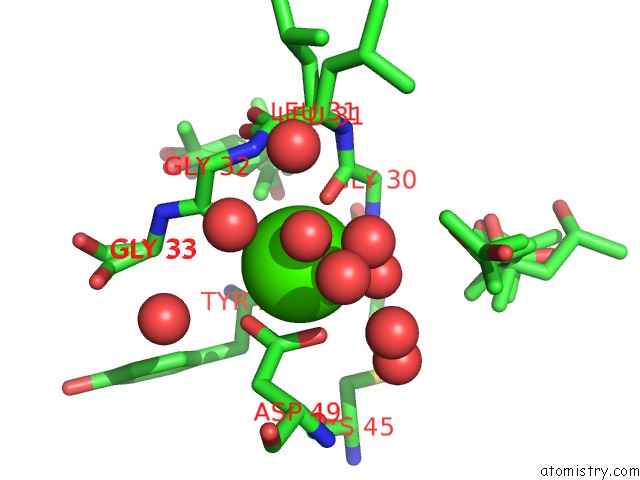
Mono view
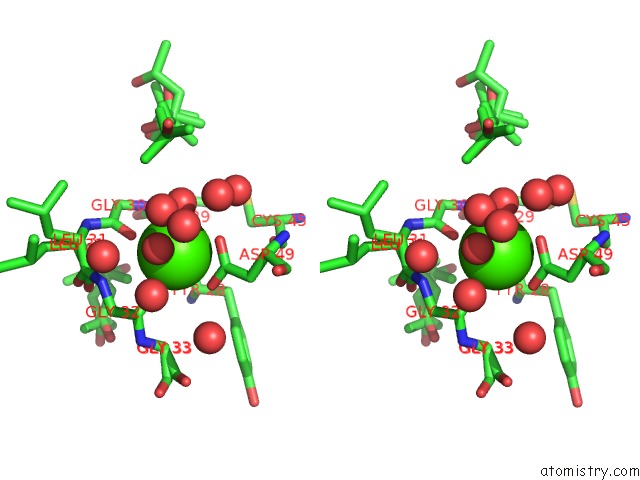
Stereo pair view
Mono view
Stereo pair view
A full contact list of Calcium with other atoms in the Ca binding
site number 1 of Atomic Resolution Structure of the Double Mutant (K53,56M) of Bovine Pancreatic Phospholipase A2 within 5.0Å range:
|
Reference:
K.Sekar,
V.Rajakannan,
D.Gayathri,
D.Velmurugan,
M.J.Poi,
M.Dauter,
Z.Dauter,
M.D.Tsai.
Atomic Resolution (0.97 A) Structure of the Triple Mutant (K53,56,121M) of Bovine Pancreatic Phospholipase A2. Acta Crystallogr.,Sect.F V. 61 3 2005.
ISSN: ESSN 1744-3091
PubMed: 16508077
DOI: 10.1107/S1744309104021748
Page generated: Tue Jul 8 04:27:35 2025
ISSN: ESSN 1744-3091
PubMed: 16508077
DOI: 10.1107/S1744309104021748
Last articles
Fe in 2YXOFe in 2YRS
Fe in 2YXC
Fe in 2YNM
Fe in 2YVJ
Fe in 2YP1
Fe in 2YU2
Fe in 2YU1
Fe in 2YQB
Fe in 2YOO