Calcium »
PDB 2b04-2bib »
2bd5 »
Calcium in PDB 2bd5: Porcine Pancreatic Elastase Complexed with Beta-Casomorphin-7 and Lys- Ser at pH 5 and Immersed in pH 9 Buffer For 30 Seconds
Enzymatic activity of Porcine Pancreatic Elastase Complexed with Beta-Casomorphin-7 and Lys- Ser at pH 5 and Immersed in pH 9 Buffer For 30 Seconds
All present enzymatic activity of Porcine Pancreatic Elastase Complexed with Beta-Casomorphin-7 and Lys- Ser at pH 5 and Immersed in pH 9 Buffer For 30 Seconds:
3.4.21.36;
3.4.21.36;
Protein crystallography data
The structure of Porcine Pancreatic Elastase Complexed with Beta-Casomorphin-7 and Lys- Ser at pH 5 and Immersed in pH 9 Buffer For 30 Seconds, PDB code: 2bd5
was solved by
B.Liu,
C.J.Schofield,
R.C.Wilmouth,
with X-Ray Crystallography technique. A brief refinement statistics is given in the table below:
| Resolution Low / High (Å) | 19.70 / 1.80 |
| Space group | P 21 21 21 |
| Cell size a, b, c (Å), α, β, γ (°) | 50.655, 57.585, 74.524, 90.00, 90.00, 90.00 |
| R / Rfree (%) | 17.4 / 21.2 |
Calcium Binding Sites:
The binding sites of Calcium atom in the Porcine Pancreatic Elastase Complexed with Beta-Casomorphin-7 and Lys- Ser at pH 5 and Immersed in pH 9 Buffer For 30 Seconds
(pdb code 2bd5). This binding sites where shown within
5.0 Angstroms radius around Calcium atom.
In total only one binding site of Calcium was determined in the Porcine Pancreatic Elastase Complexed with Beta-Casomorphin-7 and Lys- Ser at pH 5 and Immersed in pH 9 Buffer For 30 Seconds, PDB code: 2bd5:
In total only one binding site of Calcium was determined in the Porcine Pancreatic Elastase Complexed with Beta-Casomorphin-7 and Lys- Ser at pH 5 and Immersed in pH 9 Buffer For 30 Seconds, PDB code: 2bd5:
Calcium binding site 1 out of 1 in 2bd5
Go back to
Calcium binding site 1 out
of 1 in the Porcine Pancreatic Elastase Complexed with Beta-Casomorphin-7 and Lys- Ser at pH 5 and Immersed in pH 9 Buffer For 30 Seconds
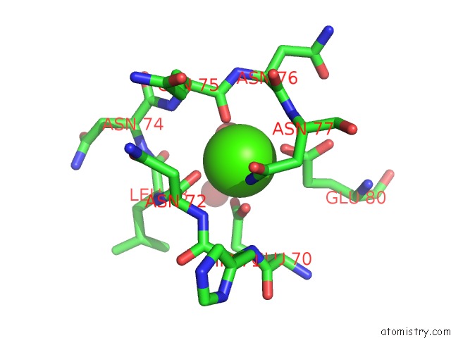
Mono view
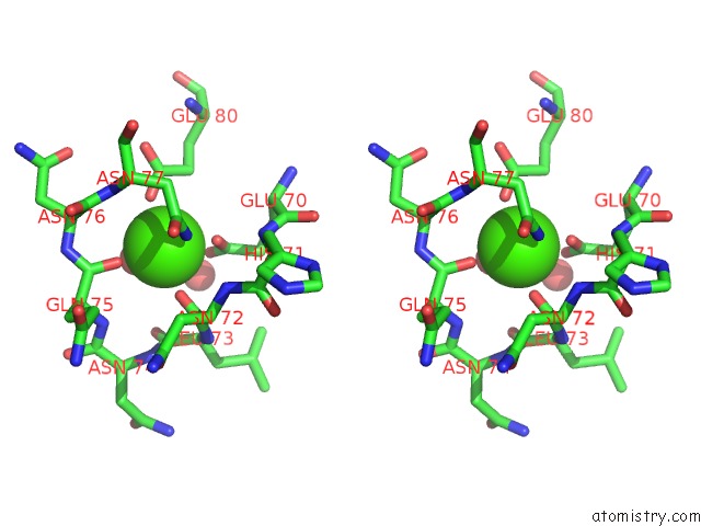
Stereo pair view
Mono view
Stereo pair view
A full contact list of Calcium with other atoms in the Ca binding
site number 1 of Porcine Pancreatic Elastase Complexed with Beta-Casomorphin-7 and Lys- Ser at pH 5 and Immersed in pH 9 Buffer For 30 Seconds within 5.0Å range:
|
Reference:
B.Liu,
C.J.Schofield,
R.C.Wilmouth.
Structural Analyses on Intermediates in Serine Protease Catalysis J.Biol.Chem. V. 281 24024 2006.
ISSN: ISSN 0021-9258
PubMed: 16754679
DOI: 10.1074/JBC.M600495200
Page generated: Fri Jul 12 09:01:15 2024
ISSN: ISSN 0021-9258
PubMed: 16754679
DOI: 10.1074/JBC.M600495200
Last articles
Zn in 9J0NZn in 9J0O
Zn in 9J0P
Zn in 9FJX
Zn in 9EKB
Zn in 9C0F
Zn in 9CAH
Zn in 9CH0
Zn in 9CH3
Zn in 9CH1