Calcium »
PDB 2yi1-2z8s »
2yih »
Calcium in PDB 2yih: Structure of A Paenibacillus Polymyxa Xyloglucanase From Gh Family 44 with Xyloglucan
Enzymatic activity of Structure of A Paenibacillus Polymyxa Xyloglucanase From Gh Family 44 with Xyloglucan
All present enzymatic activity of Structure of A Paenibacillus Polymyxa Xyloglucanase From Gh Family 44 with Xyloglucan:
3.2.1.151; 3.2.1.4;
3.2.1.151; 3.2.1.4;
Protein crystallography data
The structure of Structure of A Paenibacillus Polymyxa Xyloglucanase From Gh Family 44 with Xyloglucan, PDB code: 2yih
was solved by
A.Ariza,
J.M.Eklof,
O.Spadiut,
W.A.Offen,
S.M.Roberts,
W.Besenmatter,
E.P.Friis,
M.Skjot,
K.S.Wilson,
H.Brumer,
G.Davies,
with X-Ray Crystallography technique. A brief refinement statistics is given in the table below:
| Resolution Low / High (Å) | 41.76 / 1.70 |
| Space group | P 31 2 1 |
| Cell size a, b, c (Å), α, β, γ (°) | 83.530, 83.530, 157.140, 90.00, 90.00, 120.00 |
| R / Rfree (%) | 15.683 / 18.418 |
Other elements in 2yih:
The structure of Structure of A Paenibacillus Polymyxa Xyloglucanase From Gh Family 44 with Xyloglucan also contains other interesting chemical elements:
| Chlorine | (Cl) | 1 atom |
Calcium Binding Sites:
The binding sites of Calcium atom in the Structure of A Paenibacillus Polymyxa Xyloglucanase From Gh Family 44 with Xyloglucan
(pdb code 2yih). This binding sites where shown within
5.0 Angstroms radius around Calcium atom.
In total only one binding site of Calcium was determined in the Structure of A Paenibacillus Polymyxa Xyloglucanase From Gh Family 44 with Xyloglucan, PDB code: 2yih:
In total only one binding site of Calcium was determined in the Structure of A Paenibacillus Polymyxa Xyloglucanase From Gh Family 44 with Xyloglucan, PDB code: 2yih:
Calcium binding site 1 out of 1 in 2yih
Go back to
Calcium binding site 1 out
of 1 in the Structure of A Paenibacillus Polymyxa Xyloglucanase From Gh Family 44 with Xyloglucan
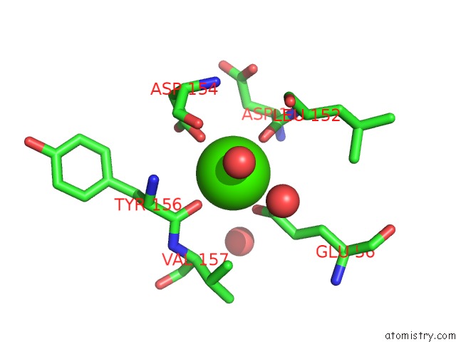
Mono view
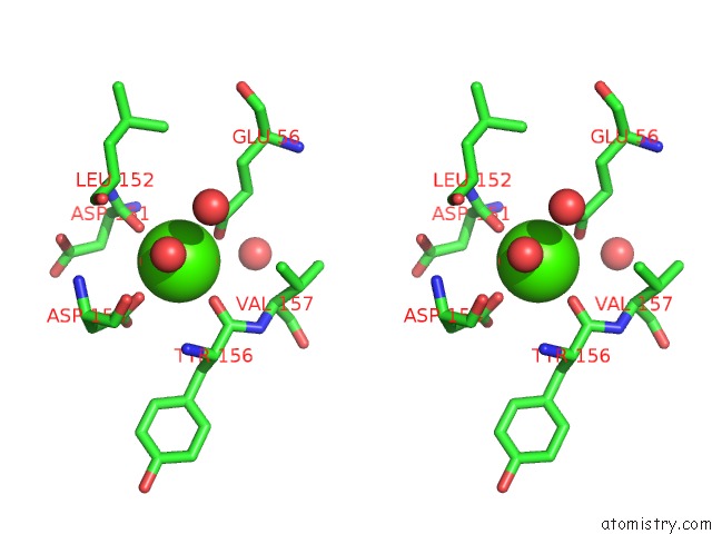
Stereo pair view
Mono view
Stereo pair view
A full contact list of Calcium with other atoms in the Ca binding
site number 1 of Structure of A Paenibacillus Polymyxa Xyloglucanase From Gh Family 44 with Xyloglucan within 5.0Å range:
|
Reference:
A.Ariza,
J.M.Eklof,
O.Spadiut,
W.A.Offen,
S.M.Roberts,
W.Besenmatter,
E.P.Friis,
M.Skjot,
K.S.Wilson,
H.Brumer,
G.Davies.
Structure and Activity of Paenibacillus Polymyxa Xyloglucanase From Glycoside Hydrolase Family 44. J.Biol.Chem. V. 286 33890 2011.
ISSN: ISSN 0021-9258
PubMed: 21795708
DOI: 10.1074/JBC.M111.262345
Page generated: Fri Jul 12 19:32:42 2024
ISSN: ISSN 0021-9258
PubMed: 21795708
DOI: 10.1074/JBC.M111.262345
Last articles
Zn in 9J0NZn in 9J0O
Zn in 9J0P
Zn in 9FJX
Zn in 9EKB
Zn in 9C0F
Zn in 9CAH
Zn in 9CH0
Zn in 9CH3
Zn in 9CH1