Calcium »
PDB 2yi1-2z8s »
2yv7 »
Calcium in PDB 2yv7: Crystal Structure of the Clic Homolog From Drosophila Melanogaster
Protein crystallography data
The structure of Crystal Structure of the Clic Homolog From Drosophila Melanogaster, PDB code: 2yv7
was solved by
S.J.Harrop,
D.R.Littler,
P.M.G.Curmi,
with X-Ray Crystallography technique. A brief refinement statistics is given in the table below:
| Resolution Low / High (Å) | 32.00 / 1.70 |
| Space group | P 21 21 21 |
| Cell size a, b, c (Å), α, β, γ (°) | 39.393, 63.451, 114.122, 90.00, 90.00, 90.00 |
| R / Rfree (%) | 21.6 / 25.4 |
Other elements in 2yv7:
The structure of Crystal Structure of the Clic Homolog From Drosophila Melanogaster also contains other interesting chemical elements:
| Iodine | (I) | 1 atom |
Calcium Binding Sites:
The binding sites of Calcium atom in the Crystal Structure of the Clic Homolog From Drosophila Melanogaster
(pdb code 2yv7). This binding sites where shown within
5.0 Angstroms radius around Calcium atom.
In total only one binding site of Calcium was determined in the Crystal Structure of the Clic Homolog From Drosophila Melanogaster, PDB code: 2yv7:
In total only one binding site of Calcium was determined in the Crystal Structure of the Clic Homolog From Drosophila Melanogaster, PDB code: 2yv7:
Calcium binding site 1 out of 1 in 2yv7
Go back to
Calcium binding site 1 out
of 1 in the Crystal Structure of the Clic Homolog From Drosophila Melanogaster
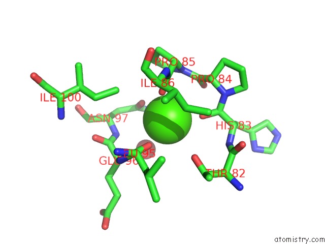
Mono view
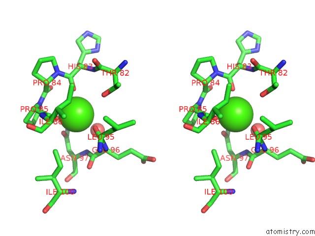
Stereo pair view
Mono view
Stereo pair view
A full contact list of Calcium with other atoms in the Ca binding
site number 1 of Crystal Structure of the Clic Homolog From Drosophila Melanogaster within 5.0Å range:
|
Reference:
D.R.Littler,
S.J.Harrop,
L.J.Brown,
G.J.Pankhurst,
A.V.Mynott,
P.Luciani,
R.A.Mandyam,
M.Mazzanti,
S.Tanda,
M.A.Berryman,
S.N.Breit,
P.M.G.Curmi.
Comparison of Vertebrate and Invertebrate Clic Proteins: the Crystal Structures of Caenorhabditis Elegans Exc-4 and Drosophila Melanogaster Dmclic Proteins V. 71 364 2007.
ISSN: ISSN 0887-3585
PubMed: 17985355
DOI: 10.1002/PROT.21704
Page generated: Tue Jul 8 09:44:15 2025
ISSN: ISSN 0887-3585
PubMed: 17985355
DOI: 10.1002/PROT.21704
Last articles
Cl in 5JS6Cl in 5JQG
Cl in 5JQS
Cl in 5JPI
Cl in 5JPH
Cl in 5JQ4
Cl in 5JQ7
Cl in 5JPV
Cl in 5JQ1
Cl in 5JOQ