Calcium »
PDB 3km6-3l4p »
3kpx »
Calcium in PDB 3kpx: Crystal Structure Analysis of Photoprotein Clytin
Enzymatic activity of Crystal Structure Analysis of Photoprotein Clytin
All present enzymatic activity of Crystal Structure Analysis of Photoprotein Clytin:
1.13.12.5;
1.13.12.5;
Protein crystallography data
The structure of Crystal Structure Analysis of Photoprotein Clytin, PDB code: 3kpx
was solved by
M.S.Titushin,
Y.Li,
G.A.Stepanyuk,
B.-C.Wang,
J.Lee,
E.S.Vysotski,
Z.-J.Liu,
with X-Ray Crystallography technique. A brief refinement statistics is given in the table below:
| Resolution Low / High (Å) | 20.31 / 1.90 |
| Space group | C 2 2 21 |
| Cell size a, b, c (Å), α, β, γ (°) | 43.392, 68.932, 115.348, 90.00, 90.00, 90.00 |
| R / Rfree (%) | 17.1 / 21.9 |
Calcium Binding Sites:
The binding sites of Calcium atom in the Crystal Structure Analysis of Photoprotein Clytin
(pdb code 3kpx). This binding sites where shown within
5.0 Angstroms radius around Calcium atom.
In total only one binding site of Calcium was determined in the Crystal Structure Analysis of Photoprotein Clytin, PDB code: 3kpx:
In total only one binding site of Calcium was determined in the Crystal Structure Analysis of Photoprotein Clytin, PDB code: 3kpx:
Calcium binding site 1 out of 1 in 3kpx
Go back to
Calcium binding site 1 out
of 1 in the Crystal Structure Analysis of Photoprotein Clytin

Mono view
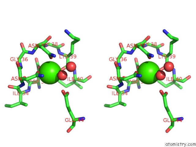
Stereo pair view
Mono view
Stereo pair view
A full contact list of Calcium with other atoms in the Ca binding
site number 1 of Crystal Structure Analysis of Photoprotein Clytin within 5.0Å range:
|
Reference:
M.S.Titushin,
Y.Feng,
G.A.Stepanyuk,
Y.Li,
S.V.Markova,
S.Golz,
B.-C.Wang,
J.Lee,
J.Wang,
E.S.Vysotski,
Z.-J.Liu.
uc(Nmr) Derived Topology of A Gfp-Photoprotein Energy Transfer Complex J.Biol.Chem. V. 285 40891 2010.
ISSN: ISSN 0021-9258
PubMed: 20926380
DOI: 10.1074/JBC.M110.133843
Page generated: Tue Jul 8 13:55:16 2025
ISSN: ISSN 0021-9258
PubMed: 20926380
DOI: 10.1074/JBC.M110.133843
Last articles
Ca in 3RDZCa in 3RBH
Ca in 3RBE
Ca in 3RBU
Ca in 3RBD
Ca in 3RB5
Ca in 3RB6
Ca in 3RB4
Ca in 3RB3
Ca in 3RB0