Calcium »
PDB 3mzo-3ngi »
3n9i »
Calcium in PDB 3n9i: Crystal Structure of Tryptophanyl-Trna Synthetase From Yersinia Pestis CO92
Enzymatic activity of Crystal Structure of Tryptophanyl-Trna Synthetase From Yersinia Pestis CO92
All present enzymatic activity of Crystal Structure of Tryptophanyl-Trna Synthetase From Yersinia Pestis CO92:
6.1.1.2;
6.1.1.2;
Protein crystallography data
The structure of Crystal Structure of Tryptophanyl-Trna Synthetase From Yersinia Pestis CO92, PDB code: 3n9i
was solved by
B.Nocek,
N.Maltseva,
L.Papazisi,
W.Anderson,
A.Joachimiak,
Center Forstructural Genomics Of Infectious Diseases (Csgid),
with X-Ray Crystallography technique. A brief refinement statistics is given in the table below:
| Resolution Low / High (Å) | 40.00 / 1.95 |
| Space group | P 21 21 21 |
| Cell size a, b, c (Å), α, β, γ (°) | 61.099, 78.150, 152.176, 90.00, 90.00, 90.00 |
| R / Rfree (%) | 16.7 / 20.6 |
Calcium Binding Sites:
The binding sites of Calcium atom in the Crystal Structure of Tryptophanyl-Trna Synthetase From Yersinia Pestis CO92
(pdb code 3n9i). This binding sites where shown within
5.0 Angstroms radius around Calcium atom.
In total only one binding site of Calcium was determined in the Crystal Structure of Tryptophanyl-Trna Synthetase From Yersinia Pestis CO92, PDB code: 3n9i:
In total only one binding site of Calcium was determined in the Crystal Structure of Tryptophanyl-Trna Synthetase From Yersinia Pestis CO92, PDB code: 3n9i:
Calcium binding site 1 out of 1 in 3n9i
Go back to
Calcium binding site 1 out
of 1 in the Crystal Structure of Tryptophanyl-Trna Synthetase From Yersinia Pestis CO92
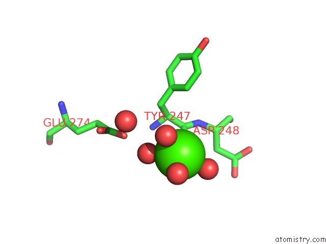
Mono view
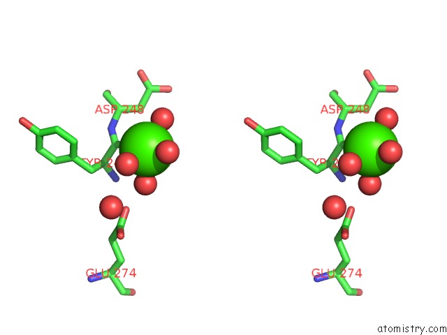
Stereo pair view
Mono view
Stereo pair view
A full contact list of Calcium with other atoms in the Ca binding
site number 1 of Crystal Structure of Tryptophanyl-Trna Synthetase From Yersinia Pestis CO92 within 5.0Å range:
|
Reference:
B.Nocek,
N.Maltseva,
L.Papazisi,
W.Anderson,
A.Joachimiak,
Center For Structural Genomics Of Infectious Diseases(Csgid).
Crystal Structure of Tryptophanyl-Trna Synthetase From Yersinia Pestis CO92 To Be Published.
Page generated: Tue Jul 8 14:49:40 2025
Last articles
Ca in 4EL8Ca in 4EFJ
Ca in 4EGY
Ca in 4EHM
Ca in 4EFS
Ca in 4EGD
Ca in 4EFZ
Ca in 4EG9
Ca in 4EE2
Ca in 4EFH