Calcium »
PDB 5jrj-5kaf »
5k8d »
Calcium in PDB 5k8d: Crystal Structure of Rfviiifc
Protein crystallography data
The structure of Crystal Structure of Rfviiifc, PDB code: 5k8d
was solved by
N.Leksa,
C.Quan,
with X-Ray Crystallography technique. A brief refinement statistics is given in the table below:
| Resolution Low / High (Å) | 37.60 / 4.19 |
| Space group | P 41 21 2 |
| Cell size a, b, c (Å), α, β, γ (°) | 136.295, 136.295, 365.098, 90.00, 90.00, 90.00 |
| R / Rfree (%) | 29.8 / 36.2 |
Other elements in 5k8d:
The structure of Crystal Structure of Rfviiifc also contains other interesting chemical elements:
| Copper | (Cu) | 2 atoms |
Calcium Binding Sites:
The binding sites of Calcium atom in the Crystal Structure of Rfviiifc
(pdb code 5k8d). This binding sites where shown within
5.0 Angstroms radius around Calcium atom.
In total 3 binding sites of Calcium where determined in the Crystal Structure of Rfviiifc, PDB code: 5k8d:
Jump to Calcium binding site number: 1; 2; 3;
In total 3 binding sites of Calcium where determined in the Crystal Structure of Rfviiifc, PDB code: 5k8d:
Jump to Calcium binding site number: 1; 2; 3;
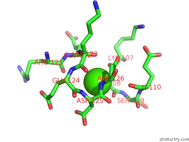
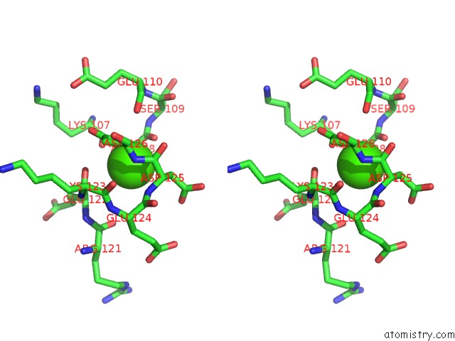
Calcium binding site 1 out of 3 in 5k8d
Go back to
Calcium binding site 1 out
of 3 in the Crystal Structure of Rfviiifc
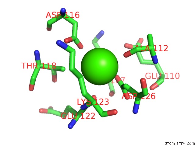
Mono view
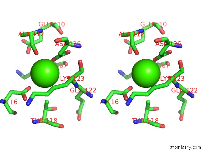
Stereo pair view
Mono view
Stereo pair view
A full contact list of Calcium with other atoms in the Ca binding
site number 1 of Crystal Structure of Rfviiifc within 5.0Å range:
|
Calcium binding site 2 out of 3 in 5k8d
Go back to
Calcium binding site 2 out
of 3 in the Crystal Structure of Rfviiifc
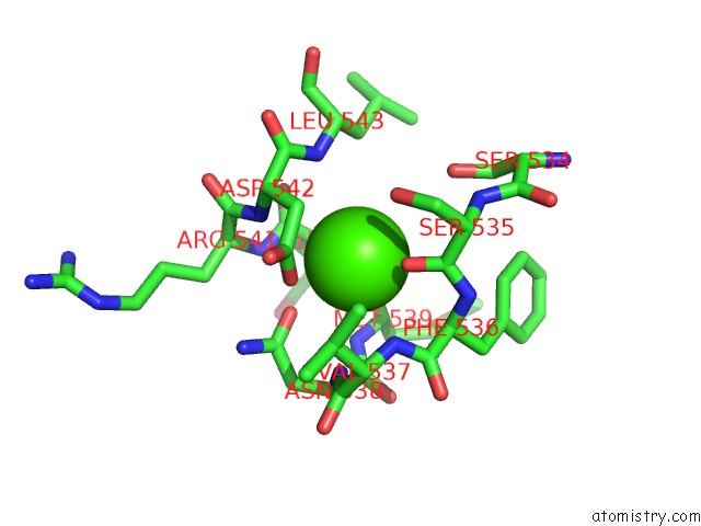
Mono view
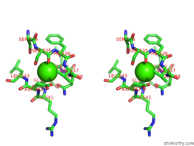
Stereo pair view
Mono view
Stereo pair view
A full contact list of Calcium with other atoms in the Ca binding
site number 2 of Crystal Structure of Rfviiifc within 5.0Å range:
|
Calcium binding site 3 out of 3 in 5k8d
Go back to
Calcium binding site 3 out
of 3 in the Crystal Structure of Rfviiifc
Mono view
Stereo pair view
Mono view
Stereo pair view
A full contact list of Calcium with other atoms in the Ca binding
site number 3 of Crystal Structure of Rfviiifc within 5.0Å range:
|
Reference:
N.C.Leksa,
P.L.Chiu,
G.M.Bou-Assaf,
C.Quan,
Z.Liu,
A.B.Goodman,
M.G.Chambers,
S.E.Tsutakawa,
M.Hammel,
R.T.Peters,
T.Walz,
J.D.Kulman.
The Structural Basis For the Functional Comparability of Factor VIII and the Long-Acting Variant Recombinant Factor VIII Fc Fusion Protein. J. Thromb. Haemost. V. 15 1167 2017.
ISSN: ESSN 1538-7836
PubMed: 28397397
DOI: 10.1111/JTH.13700
Page generated: Mon Jul 15 06:43:32 2024
ISSN: ESSN 1538-7836
PubMed: 28397397
DOI: 10.1111/JTH.13700
Last articles
Zn in 9MJ5Zn in 9HNW
Zn in 9G0L
Zn in 9FNE
Zn in 9DZN
Zn in 9E0I
Zn in 9D32
Zn in 9DAK
Zn in 8ZXC
Zn in 8ZUF