Calcium »
PDB 5jrj-5kaf »
5k8y »
Calcium in PDB 5k8y: Structure of the Mus Musclus Langerin Carbohydrate Recognition Domain
Protein crystallography data
The structure of Structure of the Mus Musclus Langerin Carbohydrate Recognition Domain, PDB code: 5k8y
was solved by
B.Loll,
J.Aretz,
C.Rademacher,
M.C.Wahl,
with X-Ray Crystallography technique. A brief refinement statistics is given in the table below:
| Resolution Low / High (Å) | 45.32 / 2.40 |
| Space group | I 2 3 |
| Cell size a, b, c (Å), α, β, γ (°) | 143.310, 143.310, 143.310, 90.00, 90.00, 90.00 |
| R / Rfree (%) | 19.6 / 23.4 |
Calcium Binding Sites:
The binding sites of Calcium atom in the Structure of the Mus Musclus Langerin Carbohydrate Recognition Domain
(pdb code 5k8y). This binding sites where shown within
5.0 Angstroms radius around Calcium atom.
In total 2 binding sites of Calcium where determined in the Structure of the Mus Musclus Langerin Carbohydrate Recognition Domain, PDB code: 5k8y:
Jump to Calcium binding site number: 1; 2;
In total 2 binding sites of Calcium where determined in the Structure of the Mus Musclus Langerin Carbohydrate Recognition Domain, PDB code: 5k8y:
Jump to Calcium binding site number: 1; 2;
Calcium binding site 1 out of 2 in 5k8y
Go back to
Calcium binding site 1 out
of 2 in the Structure of the Mus Musclus Langerin Carbohydrate Recognition Domain
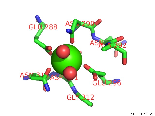
Mono view
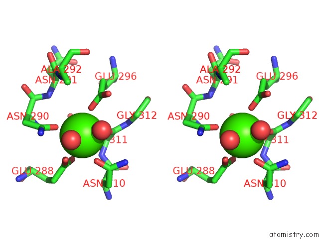
Stereo pair view
Mono view
Stereo pair view
A full contact list of Calcium with other atoms in the Ca binding
site number 1 of Structure of the Mus Musclus Langerin Carbohydrate Recognition Domain within 5.0Å range:
|
Calcium binding site 2 out of 2 in 5k8y
Go back to
Calcium binding site 2 out
of 2 in the Structure of the Mus Musclus Langerin Carbohydrate Recognition Domain
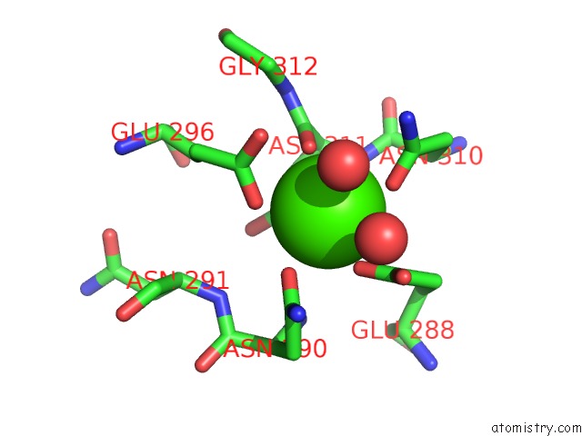
Mono view
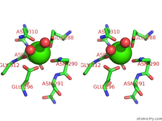
Stereo pair view
Mono view
Stereo pair view
A full contact list of Calcium with other atoms in the Ca binding
site number 2 of Structure of the Mus Musclus Langerin Carbohydrate Recognition Domain within 5.0Å range:
|
Reference:
J.Hanske,
J.Schulze,
J.Aretz,
R.Mcbride,
B.Loll,
H.Schmidt,
Y.Knirel,
W.Rabsch,
M.C.Wahl,
J.C.Paulson,
C.Rademacher.
Bacterial Polysaccharide Specificity of the Pattern Recognition Receptor Langerin Is Highly Species-Dependent. J. Biol. Chem. V. 292 862 2017.
ISSN: ESSN 1083-351X
PubMed: 27903635
DOI: 10.1074/JBC.M116.751750
Page generated: Mon Jul 15 06:45:16 2024
ISSN: ESSN 1083-351X
PubMed: 27903635
DOI: 10.1074/JBC.M116.751750
Last articles
Zn in 9MJ5Zn in 9HNW
Zn in 9G0L
Zn in 9FNE
Zn in 9DZN
Zn in 9E0I
Zn in 9D32
Zn in 9DAK
Zn in 8ZXC
Zn in 8ZUF