Calcium »
PDB 5n31-5nn9 »
5ne5 »
Calcium in PDB 5ne5: Crystal Structure of Family 47 Alpha-1,2-Mannosidase From Caulobacter K31 Strain in Complex with Kifunensine
Enzymatic activity of Crystal Structure of Family 47 Alpha-1,2-Mannosidase From Caulobacter K31 Strain in Complex with Kifunensine
All present enzymatic activity of Crystal Structure of Family 47 Alpha-1,2-Mannosidase From Caulobacter K31 Strain in Complex with Kifunensine:
3.2.1.113;
3.2.1.113;
Protein crystallography data
The structure of Crystal Structure of Family 47 Alpha-1,2-Mannosidase From Caulobacter K31 Strain in Complex with Kifunensine, PDB code: 5ne5
was solved by
A.Males,
G.J.Davies,
with X-Ray Crystallography technique. A brief refinement statistics is given in the table below:
| Resolution Low / High (Å) | 36.23 / 1.05 |
| Space group | H 3 |
| Cell size a, b, c (Å), α, β, γ (°) | 144.923, 144.923, 50.593, 90.00, 90.00, 120.00 |
| R / Rfree (%) | 14.6 / 15.2 |
Other elements in 5ne5:
The structure of Crystal Structure of Family 47 Alpha-1,2-Mannosidase From Caulobacter K31 Strain in Complex with Kifunensine also contains other interesting chemical elements:
| Sodium | (Na) | 1 atom |
Calcium Binding Sites:
The binding sites of Calcium atom in the Crystal Structure of Family 47 Alpha-1,2-Mannosidase From Caulobacter K31 Strain in Complex with Kifunensine
(pdb code 5ne5). This binding sites where shown within
5.0 Angstroms radius around Calcium atom.
In total 2 binding sites of Calcium where determined in the Crystal Structure of Family 47 Alpha-1,2-Mannosidase From Caulobacter K31 Strain in Complex with Kifunensine, PDB code: 5ne5:
Jump to Calcium binding site number: 1; 2;
In total 2 binding sites of Calcium where determined in the Crystal Structure of Family 47 Alpha-1,2-Mannosidase From Caulobacter K31 Strain in Complex with Kifunensine, PDB code: 5ne5:
Jump to Calcium binding site number: 1; 2;
Calcium binding site 1 out of 2 in 5ne5
Go back to
Calcium binding site 1 out
of 2 in the Crystal Structure of Family 47 Alpha-1,2-Mannosidase From Caulobacter K31 Strain in Complex with Kifunensine
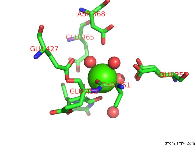
Mono view
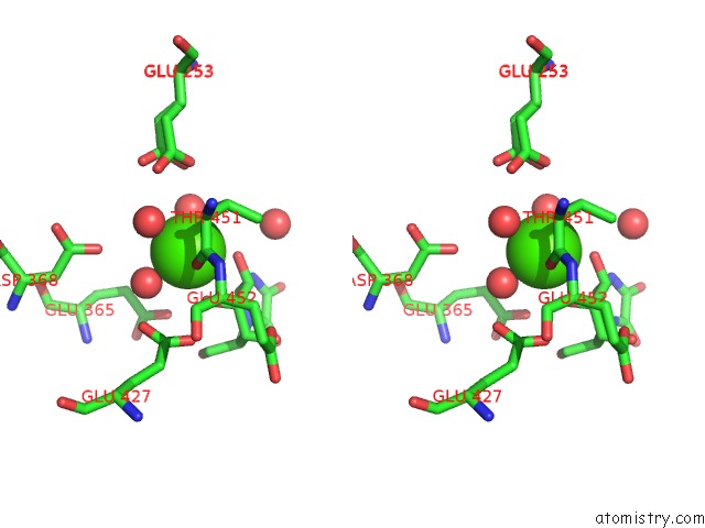
Stereo pair view
Mono view
Stereo pair view
A full contact list of Calcium with other atoms in the Ca binding
site number 1 of Crystal Structure of Family 47 Alpha-1,2-Mannosidase From Caulobacter K31 Strain in Complex with Kifunensine within 5.0Å range:
|
Calcium binding site 2 out of 2 in 5ne5
Go back to
Calcium binding site 2 out
of 2 in the Crystal Structure of Family 47 Alpha-1,2-Mannosidase From Caulobacter K31 Strain in Complex with Kifunensine
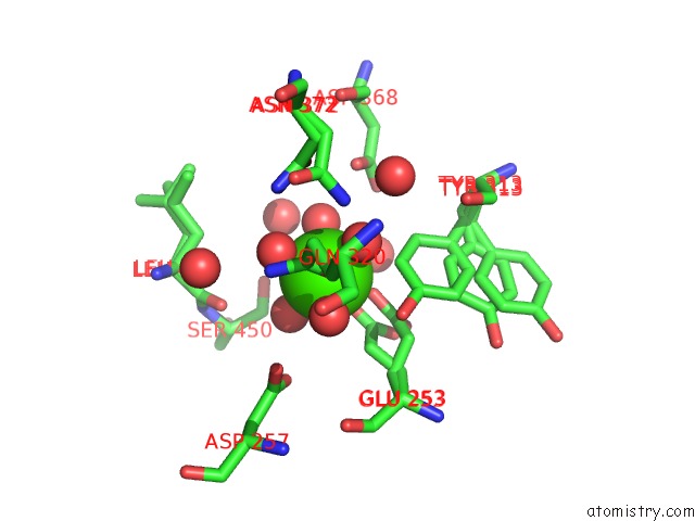
Mono view
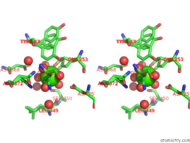
Stereo pair view
Mono view
Stereo pair view
A full contact list of Calcium with other atoms in the Ca binding
site number 2 of Crystal Structure of Family 47 Alpha-1,2-Mannosidase From Caulobacter K31 Strain in Complex with Kifunensine within 5.0Å range:
|
Reference:
A.Males,
L.Raich,
S.J.Williams,
C.Rovira,
G.J.Davies.
Conformational Analysis of the Mannosidase Inhibitor Kifunensine: A Quantum Mechanical and Structural Approach. Chembiochem V. 18 1496 2017.
ISSN: ESSN 1439-7633
PubMed: 28493500
DOI: 10.1002/CBIC.201700166
Page generated: Mon Jul 15 08:55:23 2024
ISSN: ESSN 1439-7633
PubMed: 28493500
DOI: 10.1002/CBIC.201700166
Last articles
Zn in 9J0NZn in 9J0O
Zn in 9J0P
Zn in 9FJX
Zn in 9EKB
Zn in 9C0F
Zn in 9CAH
Zn in 9CH0
Zn in 9CH3
Zn in 9CH1