Calcium »
PDB 5n31-5nn9 »
5nes »
Calcium in PDB 5nes: Discovery, Crystal Structures and Atomic Force Microscopy Study of Thioether Ligated D,L-Cyclic Antimicrobial Peptides Against Multidrug Resistant Pseudomonas Aeruginosa
Protein crystallography data
The structure of Discovery, Crystal Structures and Atomic Force Microscopy Study of Thioether Ligated D,L-Cyclic Antimicrobial Peptides Against Multidrug Resistant Pseudomonas Aeruginosa, PDB code: 5nes
was solved by
J.-L.Reymond,
T.Darbre,
A.Stocker,
W.Hong,
C.Van Delden,
T.Koehler,
A.Luscher,
R.Visini,
Y.Fu,
I.Di Bonaventura,
R.He,
with X-Ray Crystallography technique. A brief refinement statistics is given in the table below:
| Resolution Low / High (Å) | 47.78 / 1.61 |
| Space group | P 1 |
| Cell size a, b, c (Å), α, β, γ (°) | 45.201, 48.527, 52.562, 84.92, 79.98, 80.61 |
| R / Rfree (%) | 14.3 / 16.4 |
Calcium Binding Sites:
The binding sites of Calcium atom in the Discovery, Crystal Structures and Atomic Force Microscopy Study of Thioether Ligated D,L-Cyclic Antimicrobial Peptides Against Multidrug Resistant Pseudomonas Aeruginosa
(pdb code 5nes). This binding sites where shown within
5.0 Angstroms radius around Calcium atom.
In total 8 binding sites of Calcium where determined in the Discovery, Crystal Structures and Atomic Force Microscopy Study of Thioether Ligated D,L-Cyclic Antimicrobial Peptides Against Multidrug Resistant Pseudomonas Aeruginosa, PDB code: 5nes:
Jump to Calcium binding site number: 1; 2; 3; 4; 5; 6; 7; 8;
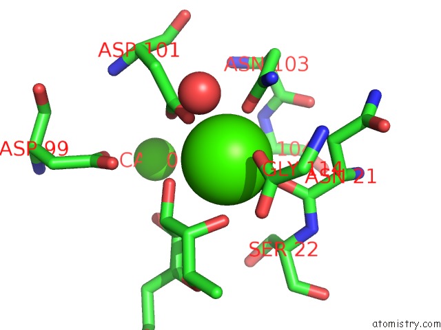
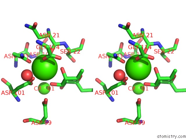
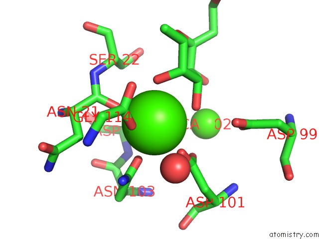
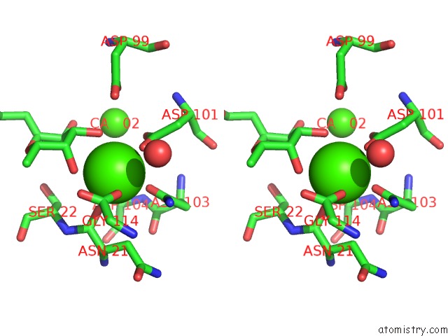
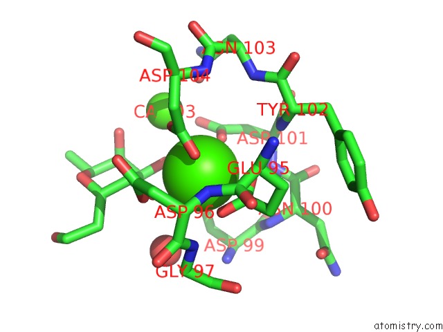
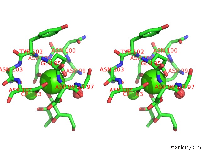
Calcium binding site 1 out of 8 in 5nes
Go back to
Calcium binding site 1 out
of 8 in the Discovery, Crystal Structures and Atomic Force Microscopy Study of Thioether Ligated D,L-Cyclic Antimicrobial Peptides Against Multidrug Resistant Pseudomonas Aeruginosa
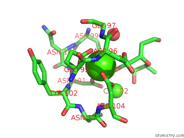
Mono view
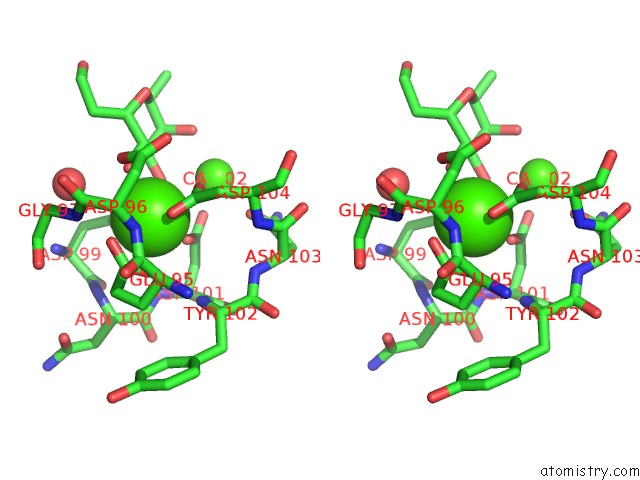
Stereo pair view
Mono view
Stereo pair view
A full contact list of Calcium with other atoms in the Ca binding
site number 1 of Discovery, Crystal Structures and Atomic Force Microscopy Study of Thioether Ligated D,L-Cyclic Antimicrobial Peptides Against Multidrug Resistant Pseudomonas Aeruginosa within 5.0Å range:
|
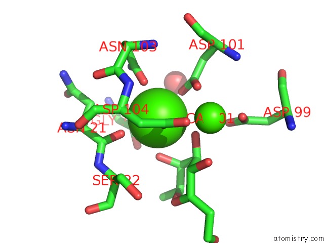
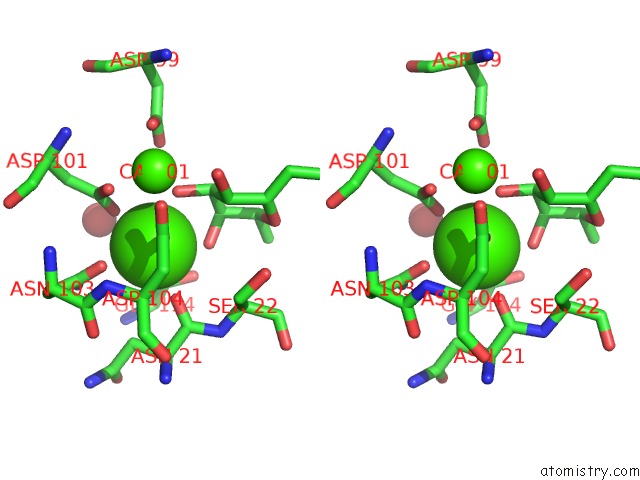
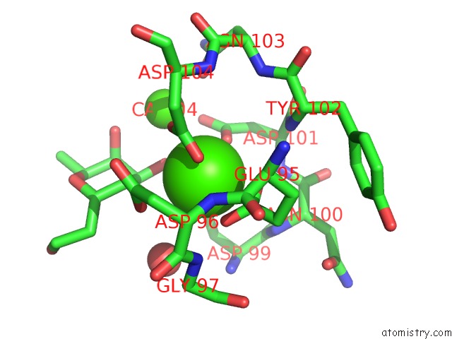
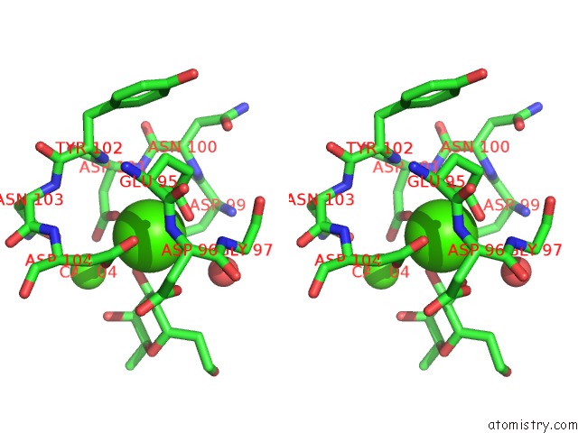
Calcium binding site 2 out of 8 in 5nes
Go back to
Calcium binding site 2 out
of 8 in the Discovery, Crystal Structures and Atomic Force Microscopy Study of Thioether Ligated D,L-Cyclic Antimicrobial Peptides Against Multidrug Resistant Pseudomonas Aeruginosa
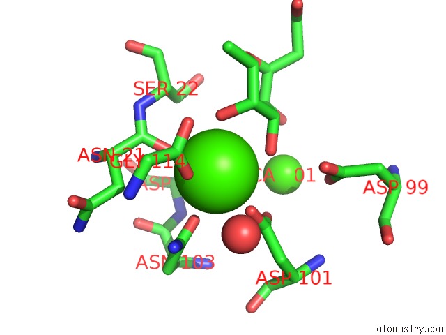
Mono view
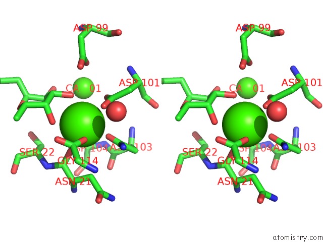
Stereo pair view
Mono view
Stereo pair view
A full contact list of Calcium with other atoms in the Ca binding
site number 2 of Discovery, Crystal Structures and Atomic Force Microscopy Study of Thioether Ligated D,L-Cyclic Antimicrobial Peptides Against Multidrug Resistant Pseudomonas Aeruginosa within 5.0Å range:
|
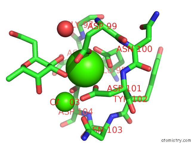
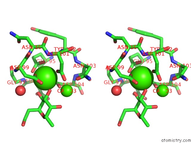
Calcium binding site 3 out of 8 in 5nes
Go back to
Calcium binding site 3 out
of 8 in the Discovery, Crystal Structures and Atomic Force Microscopy Study of Thioether Ligated D,L-Cyclic Antimicrobial Peptides Against Multidrug Resistant Pseudomonas Aeruginosa
Mono view
Stereo pair view
Mono view
Stereo pair view
A full contact list of Calcium with other atoms in the Ca binding
site number 3 of Discovery, Crystal Structures and Atomic Force Microscopy Study of Thioether Ligated D,L-Cyclic Antimicrobial Peptides Against Multidrug Resistant Pseudomonas Aeruginosa within 5.0Å range:
|
Calcium binding site 4 out of 8 in 5nes
Go back to
Calcium binding site 4 out
of 8 in the Discovery, Crystal Structures and Atomic Force Microscopy Study of Thioether Ligated D,L-Cyclic Antimicrobial Peptides Against Multidrug Resistant Pseudomonas Aeruginosa
Mono view
Stereo pair view
Mono view
Stereo pair view
A full contact list of Calcium with other atoms in the Ca binding
site number 4 of Discovery, Crystal Structures and Atomic Force Microscopy Study of Thioether Ligated D,L-Cyclic Antimicrobial Peptides Against Multidrug Resistant Pseudomonas Aeruginosa within 5.0Å range:
|
Calcium binding site 5 out of 8 in 5nes
Go back to
Calcium binding site 5 out
of 8 in the Discovery, Crystal Structures and Atomic Force Microscopy Study of Thioether Ligated D,L-Cyclic Antimicrobial Peptides Against Multidrug Resistant Pseudomonas Aeruginosa
Mono view
Stereo pair view
Mono view
Stereo pair view
A full contact list of Calcium with other atoms in the Ca binding
site number 5 of Discovery, Crystal Structures and Atomic Force Microscopy Study of Thioether Ligated D,L-Cyclic Antimicrobial Peptides Against Multidrug Resistant Pseudomonas Aeruginosa within 5.0Å range:
|
Calcium binding site 6 out of 8 in 5nes
Go back to
Calcium binding site 6 out
of 8 in the Discovery, Crystal Structures and Atomic Force Microscopy Study of Thioether Ligated D,L-Cyclic Antimicrobial Peptides Against Multidrug Resistant Pseudomonas Aeruginosa
Mono view
Stereo pair view
Mono view
Stereo pair view
A full contact list of Calcium with other atoms in the Ca binding
site number 6 of Discovery, Crystal Structures and Atomic Force Microscopy Study of Thioether Ligated D,L-Cyclic Antimicrobial Peptides Against Multidrug Resistant Pseudomonas Aeruginosa within 5.0Å range:
|
Calcium binding site 7 out of 8 in 5nes
Go back to
Calcium binding site 7 out
of 8 in the Discovery, Crystal Structures and Atomic Force Microscopy Study of Thioether Ligated D,L-Cyclic Antimicrobial Peptides Against Multidrug Resistant Pseudomonas Aeruginosa
Mono view
Stereo pair view
Mono view
Stereo pair view
A full contact list of Calcium with other atoms in the Ca binding
site number 7 of Discovery, Crystal Structures and Atomic Force Microscopy Study of Thioether Ligated D,L-Cyclic Antimicrobial Peptides Against Multidrug Resistant Pseudomonas Aeruginosa within 5.0Å range:
|
Calcium binding site 8 out of 8 in 5nes
Go back to
Calcium binding site 8 out
of 8 in the Discovery, Crystal Structures and Atomic Force Microscopy Study of Thioether Ligated D,L-Cyclic Antimicrobial Peptides Against Multidrug Resistant Pseudomonas Aeruginosa
Mono view
Stereo pair view
Mono view
Stereo pair view
A full contact list of Calcium with other atoms in the Ca binding
site number 8 of Discovery, Crystal Structures and Atomic Force Microscopy Study of Thioether Ligated D,L-Cyclic Antimicrobial Peptides Against Multidrug Resistant Pseudomonas Aeruginosa within 5.0Å range:
|
Reference:
R.He,
I.Di Bonaventura,
R.Visini,
B.H.Gan,
Y.Fu,
D.Probst,
A.Luscher,
T.Kohler,
C.Van Delden,
A.Stocker,
W.Hong,
T.Darbre,
J.L.Reymond.
Design, Crystal Structure and Atomic Force Microscopy Study of Thioether Ligated D,L-Cyclic Antimicrobial Peptides Against Multidrug Resistant Pseudomonas Aeruginosa. Chem Sci V. 8 7464 2017.
ISSN: ISSN 2041-6520
PubMed: 29163899
DOI: 10.1039/C7SC01599B
Page generated: Wed Jul 9 08:45:55 2025
ISSN: ISSN 2041-6520
PubMed: 29163899
DOI: 10.1039/C7SC01599B
Last articles
Cl in 8C61Cl in 8C6I
Cl in 8C4Q
Cl in 8C4R
Cl in 8C5Q
Cl in 8C5N
Cl in 8C5D
Cl in 8C41
Cl in 8C4N
Cl in 8C4O