Calcium »
PDB 5n31-5nn9 »
5ngy »
Calcium in PDB 5ngy: Crystal Structure of Leuconostoc Citreum Nrrl B-1299 Dextransucrase Dsr-M
Protein crystallography data
The structure of Crystal Structure of Leuconostoc Citreum Nrrl B-1299 Dextransucrase Dsr-M, PDB code: 5ngy
was solved by
M.Claverie,
G.Cioci,
M.Remaud-Simeon,
C.Moulis,
S.Tranier,
M.Vuillemin,
with X-Ray Crystallography technique. A brief refinement statistics is given in the table below:
| Resolution Low / High (Å) | 50.00 / 3.70 |
| Space group | P 21 21 21 |
| Cell size a, b, c (Å), α, β, γ (°) | 105.869, 128.810, 234.518, 90.00, 90.00, 90.00 |
| R / Rfree (%) | 19.2 / 23.6 |
Other elements in 5ngy:
The structure of Crystal Structure of Leuconostoc Citreum Nrrl B-1299 Dextransucrase Dsr-M also contains other interesting chemical elements:
| Praseodymium | (Pr) | 6 atoms |
Calcium Binding Sites:
The binding sites of Calcium atom in the Crystal Structure of Leuconostoc Citreum Nrrl B-1299 Dextransucrase Dsr-M
(pdb code 5ngy). This binding sites where shown within
5.0 Angstroms radius around Calcium atom.
In total 2 binding sites of Calcium where determined in the Crystal Structure of Leuconostoc Citreum Nrrl B-1299 Dextransucrase Dsr-M, PDB code: 5ngy:
Jump to Calcium binding site number: 1; 2;
In total 2 binding sites of Calcium where determined in the Crystal Structure of Leuconostoc Citreum Nrrl B-1299 Dextransucrase Dsr-M, PDB code: 5ngy:
Jump to Calcium binding site number: 1; 2;
Calcium binding site 1 out of 2 in 5ngy
Go back to
Calcium binding site 1 out
of 2 in the Crystal Structure of Leuconostoc Citreum Nrrl B-1299 Dextransucrase Dsr-M
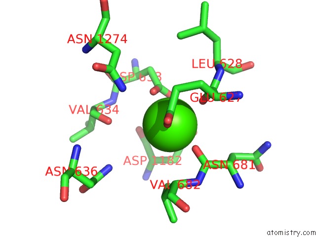
Mono view
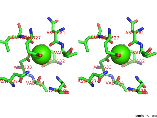
Stereo pair view
Mono view
Stereo pair view
A full contact list of Calcium with other atoms in the Ca binding
site number 1 of Crystal Structure of Leuconostoc Citreum Nrrl B-1299 Dextransucrase Dsr-M within 5.0Å range:
|
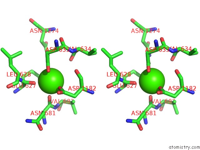
Calcium binding site 2 out of 2 in 5ngy
Go back to
Calcium binding site 2 out
of 2 in the Crystal Structure of Leuconostoc Citreum Nrrl B-1299 Dextransucrase Dsr-M
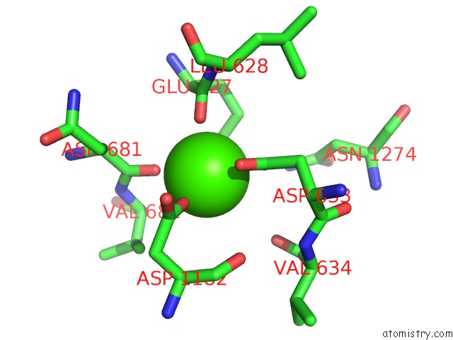
Mono view
Stereo pair view
Mono view
Stereo pair view
A full contact list of Calcium with other atoms in the Ca binding
site number 2 of Crystal Structure of Leuconostoc Citreum Nrrl B-1299 Dextransucrase Dsr-M within 5.0Å range:
|
Reference:
M.Claverie,
G.Cioci,
M.Vuillemin,
N.Monties,
P.Roblin,
G.Lippens,
M.Remaud-Simeon,
C.Moulis.
Investigations on the Determinants Responsible For Low Molar Mass Dextran Formation By Dsr-M Dextransucrase Acs Catalysis 2017.
ISSN: ESSN 2155-5435
DOI: 10.1021/ACSCATAL.7B02182
Page generated: Wed Jul 9 08:52:28 2025
ISSN: ESSN 2155-5435
DOI: 10.1021/ACSCATAL.7B02182
Last articles
Fe in 2YXOFe in 2YRS
Fe in 2YXC
Fe in 2YNM
Fe in 2YVJ
Fe in 2YP1
Fe in 2YU2
Fe in 2YU1
Fe in 2YQB
Fe in 2YOO