Calcium »
PDB 5s5v-5szl »
5sv0 »
Calcium in PDB 5sv0: Structure of the Exbb/Exbd Complex From E. Coli at pH 7.0
Protein crystallography data
The structure of Structure of the Exbb/Exbd Complex From E. Coli at pH 7.0, PDB code: 5sv0
was solved by
H.Celia,
I.Botos,
R.Lloubes,
S.K.Buchanan,
N.Noinaj,
with X-Ray Crystallography technique. A brief refinement statistics is given in the table below:
| Resolution Low / High (Å) | 46.00 / 2.60 |
| Space group | P 1 21 1 |
| Cell size a, b, c (Å), α, β, γ (°) | 137.166, 104.779, 149.364, 90.00, 91.77, 90.00 |
| R / Rfree (%) | 21.1 / 25.8 |
Calcium Binding Sites:
The binding sites of Calcium atom in the Structure of the Exbb/Exbd Complex From E. Coli at pH 7.0
(pdb code 5sv0). This binding sites where shown within
5.0 Angstroms radius around Calcium atom.
In total 2 binding sites of Calcium where determined in the Structure of the Exbb/Exbd Complex From E. Coli at pH 7.0, PDB code: 5sv0:
Jump to Calcium binding site number: 1; 2;
In total 2 binding sites of Calcium where determined in the Structure of the Exbb/Exbd Complex From E. Coli at pH 7.0, PDB code: 5sv0:
Jump to Calcium binding site number: 1; 2;
Calcium binding site 1 out of 2 in 5sv0
Go back to
Calcium binding site 1 out
of 2 in the Structure of the Exbb/Exbd Complex From E. Coli at pH 7.0
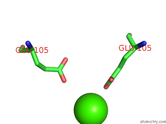
Mono view
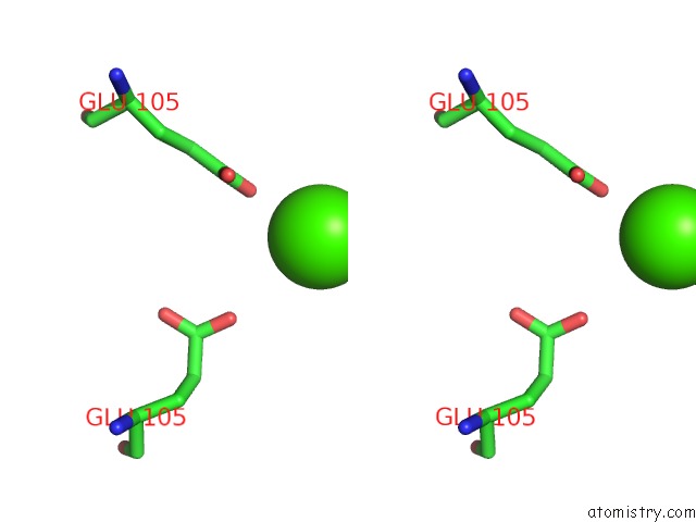
Stereo pair view
Mono view
Stereo pair view
A full contact list of Calcium with other atoms in the Ca binding
site number 1 of Structure of the Exbb/Exbd Complex From E. Coli at pH 7.0 within 5.0Å range:
|
Calcium binding site 2 out of 2 in 5sv0
Go back to
Calcium binding site 2 out
of 2 in the Structure of the Exbb/Exbd Complex From E. Coli at pH 7.0
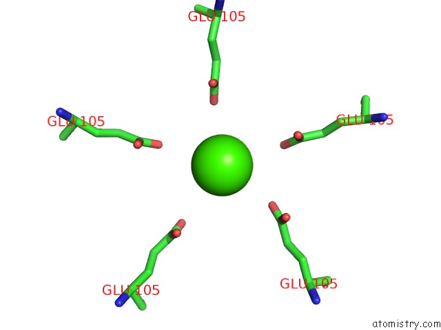
Mono view
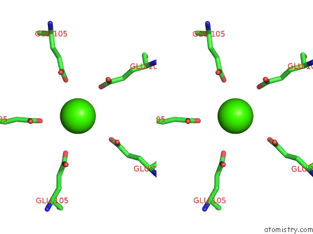
Stereo pair view
Mono view
Stereo pair view
A full contact list of Calcium with other atoms in the Ca binding
site number 2 of Structure of the Exbb/Exbd Complex From E. Coli at pH 7.0 within 5.0Å range:
|
Reference:
H.Celia,
N.Noinaj,
S.D.Zakharov,
E.Bordignon,
I.Botos,
M.Santamaria,
T.J.Barnard,
W.A.Cramer,
R.Lloubes,
S.K.Buchanan.
Structural Insight Into the Role of the Ton Complex in Energy Transduction. Nature V. 538 60 2016.
ISSN: ESSN 1476-4687
PubMed: 27654919
DOI: 10.1038/NATURE19757
Page generated: Wed Jul 9 09:50:04 2025
ISSN: ESSN 1476-4687
PubMed: 27654919
DOI: 10.1038/NATURE19757
Last articles
Cl in 5HQJCl in 5HQC
Cl in 5HQH
Cl in 5HQ1
Cl in 5HQ4
Cl in 5HML
Cl in 5HM8
Cl in 5HPO
Cl in 5HPU
Cl in 5HPP