Calcium »
PDB 5s5v-5szl »
5swi »
Calcium in PDB 5swi: Crystal Structure of SPGH92 in Complex with Mannose
Protein crystallography data
The structure of Crystal Structure of SPGH92 in Complex with Mannose, PDB code: 5swi
was solved by
S.Shapiro-Ward,
A.B.Boraston,
with X-Ray Crystallography technique. A brief refinement statistics is given in the table below:
| Resolution Low / High (Å) | 127.76 / 2.15 |
| Space group | P 21 21 21 |
| Cell size a, b, c (Å), α, β, γ (°) | 130.003, 161.711, 208.404, 90.00, 90.00, 90.00 |
| R / Rfree (%) | 19.7 / 22.1 |
Calcium Binding Sites:
The binding sites of Calcium atom in the Crystal Structure of SPGH92 in Complex with Mannose
(pdb code 5swi). This binding sites where shown within
5.0 Angstroms radius around Calcium atom.
In total 4 binding sites of Calcium where determined in the Crystal Structure of SPGH92 in Complex with Mannose, PDB code: 5swi:
Jump to Calcium binding site number: 1; 2; 3; 4;
In total 4 binding sites of Calcium where determined in the Crystal Structure of SPGH92 in Complex with Mannose, PDB code: 5swi:
Jump to Calcium binding site number: 1; 2; 3; 4;
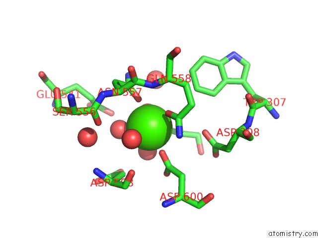
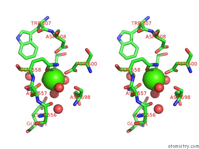
Calcium binding site 1 out of 4 in 5swi
Go back to
Calcium binding site 1 out
of 4 in the Crystal Structure of SPGH92 in Complex with Mannose
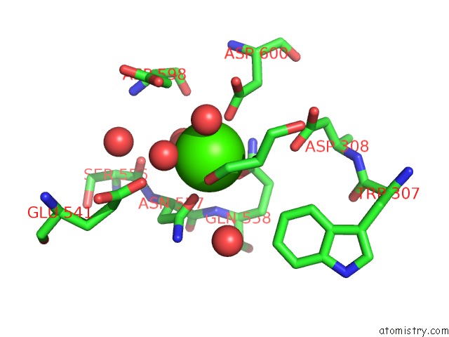
Mono view
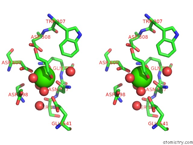
Stereo pair view
Mono view
Stereo pair view
A full contact list of Calcium with other atoms in the Ca binding
site number 1 of Crystal Structure of SPGH92 in Complex with Mannose within 5.0Å range:
|
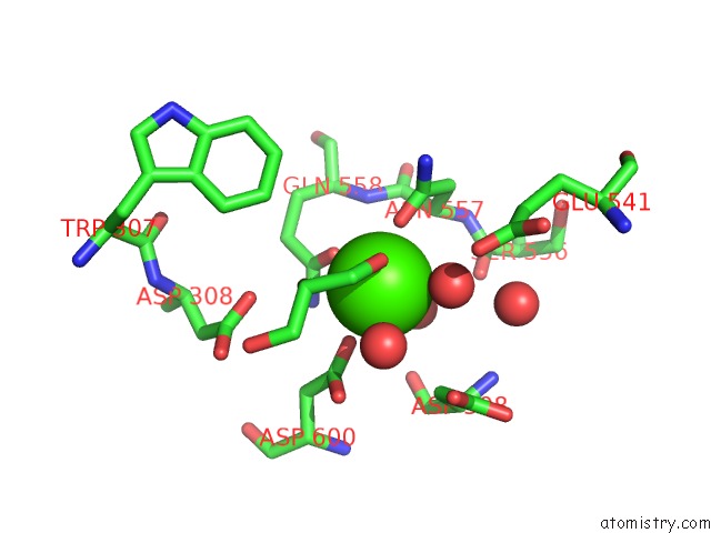
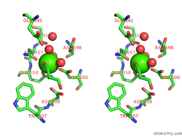
Calcium binding site 2 out of 4 in 5swi
Go back to
Calcium binding site 2 out
of 4 in the Crystal Structure of SPGH92 in Complex with Mannose
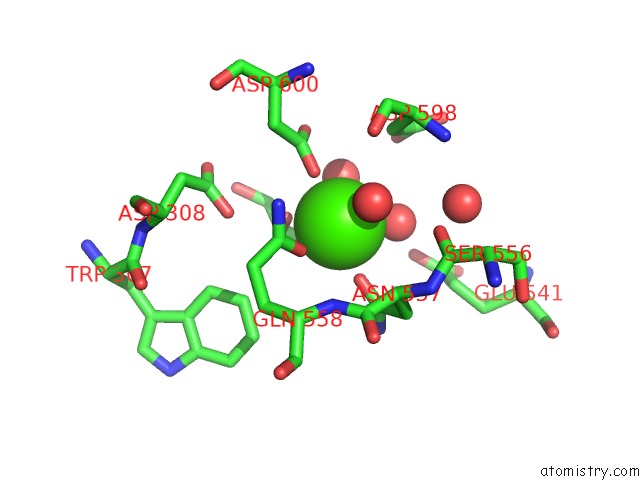
Mono view
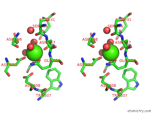
Stereo pair view
Mono view
Stereo pair view
A full contact list of Calcium with other atoms in the Ca binding
site number 2 of Crystal Structure of SPGH92 in Complex with Mannose within 5.0Å range:
|
Calcium binding site 3 out of 4 in 5swi
Go back to
Calcium binding site 3 out
of 4 in the Crystal Structure of SPGH92 in Complex with Mannose
Mono view
Stereo pair view
Mono view
Stereo pair view
A full contact list of Calcium with other atoms in the Ca binding
site number 3 of Crystal Structure of SPGH92 in Complex with Mannose within 5.0Å range:
|
Calcium binding site 4 out of 4 in 5swi
Go back to
Calcium binding site 4 out
of 4 in the Crystal Structure of SPGH92 in Complex with Mannose
Mono view
Stereo pair view
Mono view
Stereo pair view
A full contact list of Calcium with other atoms in the Ca binding
site number 4 of Crystal Structure of SPGH92 in Complex with Mannose within 5.0Å range:
|
Reference:
M.Robb,
J.K.Hobbs,
S.A.Woodiga,
S.Shapiro-Ward,
M.D.Suits,
N.Mcgregor,
H.Brumer,
H.Yesilkaya,
S.J.King,
A.B.Boraston.
Molecular Characterization of N-Glycan Degradation and Transport in Streptococcus Pneumoniae and Its Contribution to Virulence. Plos Pathog. V. 13 06090 2017.
ISSN: ESSN 1553-7374
PubMed: 28056108
DOI: 10.1371/JOURNAL.PPAT.1006090
Page generated: Wed Jul 9 09:50:20 2025
ISSN: ESSN 1553-7374
PubMed: 28056108
DOI: 10.1371/JOURNAL.PPAT.1006090
Last articles
F in 7L4NF in 7L4M
F in 7L4T
F in 7L4K
F in 7L4F
F in 7L26
F in 7L4H
F in 7L4C
F in 7L24
F in 7L25