Calcium »
PDB 5wn4-5wzv »
5wpr »
Calcium in PDB 5wpr: Crystal Structure HPIC1 in C2 Space Group
Protein crystallography data
The structure of Crystal Structure HPIC1 in C2 Space Group, PDB code: 5wpr
was solved by
S.A.Newmister,
S.Li,
M.Garcia-Borras,
J.N.Sanders,
S.Yang,
A.N.Lowell,
F.Yu,
J.L.Smith,
R.M.Williams,
K.N.Houk,
D.H.Sherman,
with X-Ray Crystallography technique. A brief refinement statistics is given in the table below:
| Resolution Low / High (Å) | 45.11 / 1.49 |
| Space group | C 1 2 1 |
| Cell size a, b, c (Å), α, β, γ (°) | 113.846, 49.522, 53.135, 90.00, 110.51, 90.00 |
| R / Rfree (%) | 16.3 / 18.3 |
Calcium Binding Sites:
The binding sites of Calcium atom in the Crystal Structure HPIC1 in C2 Space Group
(pdb code 5wpr). This binding sites where shown within
5.0 Angstroms radius around Calcium atom.
In total 2 binding sites of Calcium where determined in the Crystal Structure HPIC1 in C2 Space Group, PDB code: 5wpr:
Jump to Calcium binding site number: 1; 2;
In total 2 binding sites of Calcium where determined in the Crystal Structure HPIC1 in C2 Space Group, PDB code: 5wpr:
Jump to Calcium binding site number: 1; 2;
Calcium binding site 1 out of 2 in 5wpr
Go back to
Calcium binding site 1 out
of 2 in the Crystal Structure HPIC1 in C2 Space Group
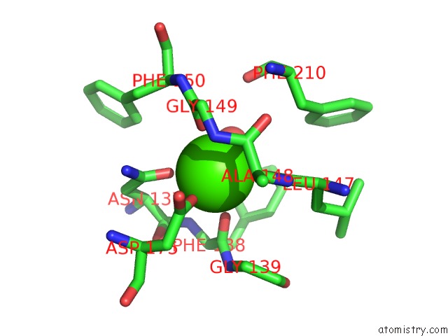
Mono view
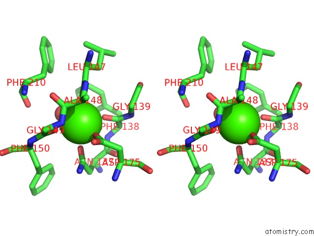
Stereo pair view
Mono view
Stereo pair view
A full contact list of Calcium with other atoms in the Ca binding
site number 1 of Crystal Structure HPIC1 in C2 Space Group within 5.0Å range:
|
Calcium binding site 2 out of 2 in 5wpr
Go back to
Calcium binding site 2 out
of 2 in the Crystal Structure HPIC1 in C2 Space Group
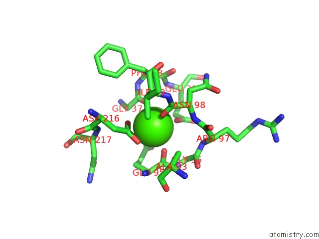
Mono view
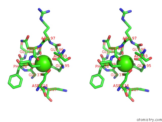
Stereo pair view
Mono view
Stereo pair view
A full contact list of Calcium with other atoms in the Ca binding
site number 2 of Crystal Structure HPIC1 in C2 Space Group within 5.0Å range:
|
Reference:
S.A.Newmister,
S.Li,
M.Garcia-Borras,
J.N.Sanders,
S.Yang,
A.N.Lowell,
F.Yu,
J.L.Smith,
R.M.Williams,
K.N.Houk,
D.H.Sherman.
Structural Basis of the Cope Rearrangement and Cyclization in Hapalindole Biogenesis. Nat. Chem. Biol. V. 14 345 2018.
ISSN: ESSN 1552-4469
PubMed: 29531360
DOI: 10.1038/S41589-018-0003-X
Page generated: Wed Jul 9 11:20:02 2025
ISSN: ESSN 1552-4469
PubMed: 29531360
DOI: 10.1038/S41589-018-0003-X
Last articles
Ca in 7PKBCa in 7PI3
Ca in 7PJM
Ca in 7PHW
Ca in 7PIV
Ca in 7PI0
Ca in 7PIU
Ca in 7PC9
Ca in 7PH1
Ca in 7PEO