Calcium »
PDB 6e54-6ela »
6eax »
Calcium in PDB 6eax: Crystallographic Structure of the Cyclic Hexapeptide Derived From the Btci Inhibitor Bound to Beta-Trypsin in Space Group P 21 21 21
Enzymatic activity of Crystallographic Structure of the Cyclic Hexapeptide Derived From the Btci Inhibitor Bound to Beta-Trypsin in Space Group P 21 21 21
All present enzymatic activity of Crystallographic Structure of the Cyclic Hexapeptide Derived From the Btci Inhibitor Bound to Beta-Trypsin in Space Group P 21 21 21:
3.4.21.4;
3.4.21.4;
Protein crystallography data
The structure of Crystallographic Structure of the Cyclic Hexapeptide Derived From the Btci Inhibitor Bound to Beta-Trypsin in Space Group P 21 21 21, PDB code: 6eax
was solved by
J.C.Fernandes,
N.F.Valadares,
S.M.Freitas,
J.A.R.G.Barbosa,
with X-Ray Crystallography technique. A brief refinement statistics is given in the table below:
| Resolution Low / High (Å) | 23.33 / 1.19 |
| Space group | P 21 21 21 |
| Cell size a, b, c (Å), α, β, γ (°) | 61.585, 63.544, 68.754, 90.00, 90.00, 90.00 |
| R / Rfree (%) | 14.3 / 15.6 |
Calcium Binding Sites:
The binding sites of Calcium atom in the Crystallographic Structure of the Cyclic Hexapeptide Derived From the Btci Inhibitor Bound to Beta-Trypsin in Space Group P 21 21 21
(pdb code 6eax). This binding sites where shown within
5.0 Angstroms radius around Calcium atom.
In total only one binding site of Calcium was determined in the Crystallographic Structure of the Cyclic Hexapeptide Derived From the Btci Inhibitor Bound to Beta-Trypsin in Space Group P 21 21 21, PDB code: 6eax:
In total only one binding site of Calcium was determined in the Crystallographic Structure of the Cyclic Hexapeptide Derived From the Btci Inhibitor Bound to Beta-Trypsin in Space Group P 21 21 21, PDB code: 6eax:
Calcium binding site 1 out of 1 in 6eax
Go back to
Calcium binding site 1 out
of 1 in the Crystallographic Structure of the Cyclic Hexapeptide Derived From the Btci Inhibitor Bound to Beta-Trypsin in Space Group P 21 21 21
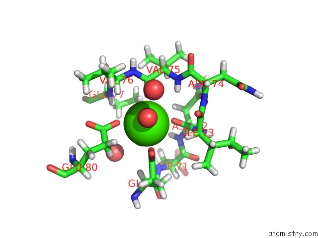
Mono view
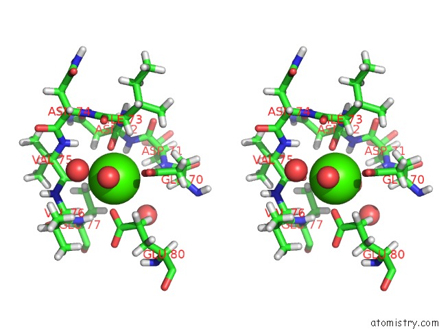
Stereo pair view
Mono view
Stereo pair view
A full contact list of Calcium with other atoms in the Ca binding
site number 1 of Crystallographic Structure of the Cyclic Hexapeptide Derived From the Btci Inhibitor Bound to Beta-Trypsin in Space Group P 21 21 21 within 5.0Å range:
|
Reference:
J.F.Fenandes,
N.F.Valadares,
S.M.Freitas,
J.A.R.G.Barbosa.
Crystallographic Structure of the Cyclic Hexapeptide Derived From the Btci Inhibitor Bound to Beta-Trypsin in Space Group P 21 21 21 To Be Published.
Page generated: Wed Jul 9 13:37:32 2025
Last articles
Ca in 7ET8Ca in 7ESR
Ca in 7ESY
Ca in 7EST
Ca in 7EMJ
Ca in 7ESK
Ca in 7ESI
Ca in 7ERJ
Ca in 7ERH
Ca in 7EQ7