Calcium »
PDB 6f30-6fi4 »
6f7l »
Calcium in PDB 6f7l: Crystal Structure of Lkce R326Q Mutant in Complex with Its Substrate
Protein crystallography data
The structure of Crystal Structure of Lkce R326Q Mutant in Complex with Its Substrate, PDB code: 6f7l
was solved by
J.Dorival,
F.Risser,
C.Jacob,
S.Collin,
G.Drager,
A.Kirschning,
C.Paris,
B.Chagot,
A.Gruez,
K.J.Weissman,
with X-Ray Crystallography technique. A brief refinement statistics is given in the table below:
| Resolution Low / High (Å) | 48.99 / 2.50 |
| Space group | P 41 21 2 |
| Cell size a, b, c (Å), α, β, γ (°) | 125.554, 125.554, 156.655, 90.00, 90.00, 90.00 |
| R / Rfree (%) | 20.6 / 24.4 |
Calcium Binding Sites:
The binding sites of Calcium atom in the Crystal Structure of Lkce R326Q Mutant in Complex with Its Substrate
(pdb code 6f7l). This binding sites where shown within
5.0 Angstroms radius around Calcium atom.
In total 3 binding sites of Calcium where determined in the Crystal Structure of Lkce R326Q Mutant in Complex with Its Substrate, PDB code: 6f7l:
Jump to Calcium binding site number: 1; 2; 3;
In total 3 binding sites of Calcium where determined in the Crystal Structure of Lkce R326Q Mutant in Complex with Its Substrate, PDB code: 6f7l:
Jump to Calcium binding site number: 1; 2; 3;
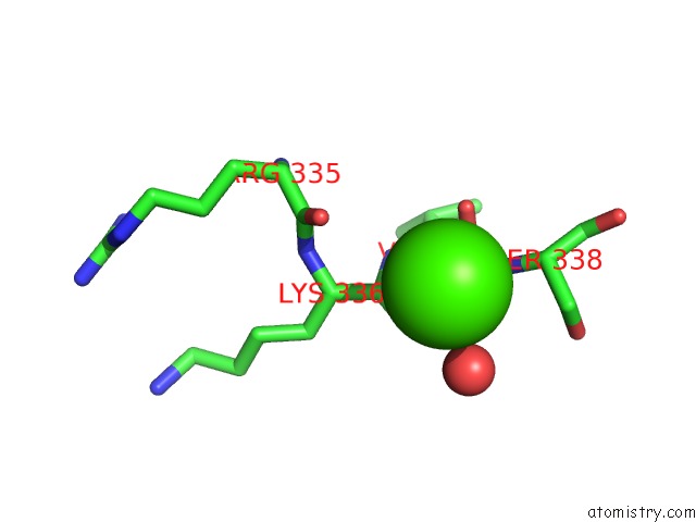
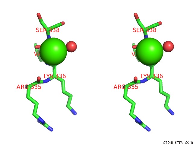
Calcium binding site 1 out of 3 in 6f7l
Go back to
Calcium binding site 1 out
of 3 in the Crystal Structure of Lkce R326Q Mutant in Complex with Its Substrate
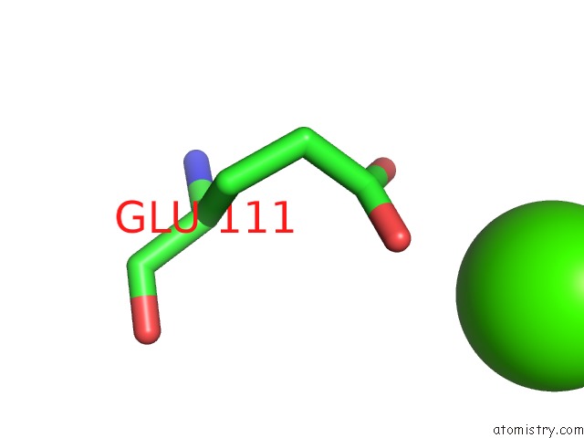
Mono view
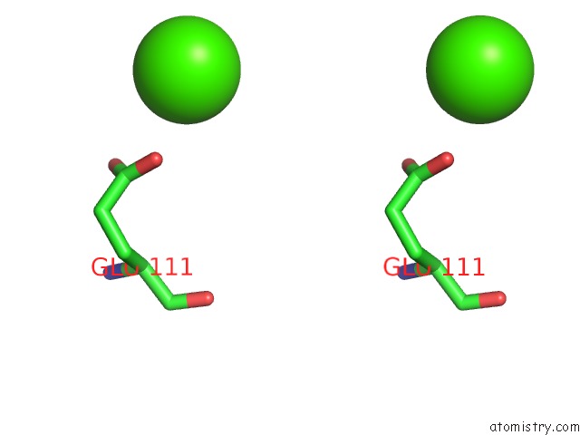
Stereo pair view
Mono view
Stereo pair view
A full contact list of Calcium with other atoms in the Ca binding
site number 1 of Crystal Structure of Lkce R326Q Mutant in Complex with Its Substrate within 5.0Å range:
|
Calcium binding site 2 out of 3 in 6f7l
Go back to
Calcium binding site 2 out
of 3 in the Crystal Structure of Lkce R326Q Mutant in Complex with Its Substrate
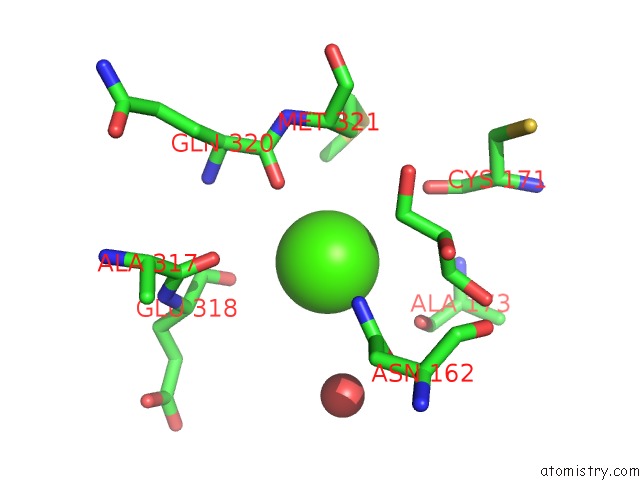
Mono view
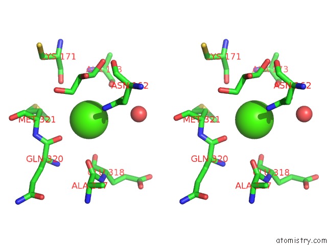
Stereo pair view
Mono view
Stereo pair view
A full contact list of Calcium with other atoms in the Ca binding
site number 2 of Crystal Structure of Lkce R326Q Mutant in Complex with Its Substrate within 5.0Å range:
|
Calcium binding site 3 out of 3 in 6f7l
Go back to
Calcium binding site 3 out
of 3 in the Crystal Structure of Lkce R326Q Mutant in Complex with Its Substrate
Mono view
Stereo pair view
Mono view
Stereo pair view
A full contact list of Calcium with other atoms in the Ca binding
site number 3 of Crystal Structure of Lkce R326Q Mutant in Complex with Its Substrate within 5.0Å range:
|
Reference:
J.Dorival,
F.Risser,
C.Jacob,
S.Collin,
G.Drager,
C.Paris,
B.Chagot,
A.Kirschning,
A.Gruez,
K.J.Weissman.
Insights Into A Dual Function Amide Oxidase/Macrocyclase From Lankacidin Biosynthesis. Nat Commun V. 9 3998 2018.
ISSN: ESSN 2041-1723
PubMed: 30266997
DOI: 10.1038/S41467-018-06323-W
Page generated: Wed Jul 9 14:07:47 2025
ISSN: ESSN 2041-1723
PubMed: 30266997
DOI: 10.1038/S41467-018-06323-W
Last articles
Ca in 7F58Ca in 7F55
Ca in 7F54
Ca in 7F4Y
Ca in 7F53
Ca in 7F4I
Ca in 7F4H
Ca in 7F4D
Ca in 7F4F
Ca in 7EV1