Calcium »
PDB 6fzw-6guy »
6gh8 »
Calcium in PDB 6gh8: Crystal Structure of GP1 Domain of Lujo Virus in Complex with the First Cub Domain of Neuropilin-2
Protein crystallography data
The structure of Crystal Structure of GP1 Domain of Lujo Virus in Complex with the First Cub Domain of Neuropilin-2, PDB code: 6gh8
was solved by
H.Cohen-Dvashi,
I.Kilimnik,
R.Diskin,
with X-Ray Crystallography technique. A brief refinement statistics is given in the table below:
| Resolution Low / High (Å) | 72.00 / 2.44 |
| Space group | P 21 21 2 |
| Cell size a, b, c (Å), α, β, γ (°) | 141.808, 59.294, 83.567, 90.00, 90.00, 90.00 |
| R / Rfree (%) | 26.6 / 31.2 |
Calcium Binding Sites:
The binding sites of Calcium atom in the Crystal Structure of GP1 Domain of Lujo Virus in Complex with the First Cub Domain of Neuropilin-2
(pdb code 6gh8). This binding sites where shown within
5.0 Angstroms radius around Calcium atom.
In total 2 binding sites of Calcium where determined in the Crystal Structure of GP1 Domain of Lujo Virus in Complex with the First Cub Domain of Neuropilin-2, PDB code: 6gh8:
Jump to Calcium binding site number: 1; 2;
In total 2 binding sites of Calcium where determined in the Crystal Structure of GP1 Domain of Lujo Virus in Complex with the First Cub Domain of Neuropilin-2, PDB code: 6gh8:
Jump to Calcium binding site number: 1; 2;
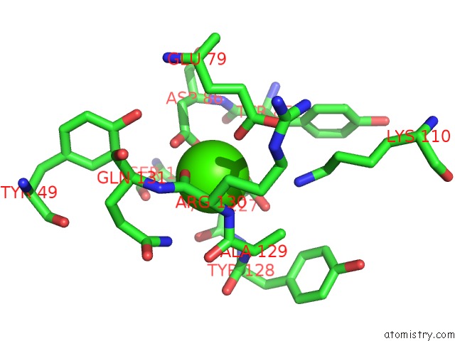
Calcium binding site 1 out of 2 in 6gh8
Go back to
Calcium binding site 1 out
of 2 in the Crystal Structure of GP1 Domain of Lujo Virus in Complex with the First Cub Domain of Neuropilin-2

Mono view

Stereo pair view
Mono view
Stereo pair view
A full contact list of Calcium with other atoms in the Ca binding
site number 1 of Crystal Structure of GP1 Domain of Lujo Virus in Complex with the First Cub Domain of Neuropilin-2 within 5.0Å range:
|
Calcium binding site 2 out of 2 in 6gh8
Go back to
Calcium binding site 2 out
of 2 in the Crystal Structure of GP1 Domain of Lujo Virus in Complex with the First Cub Domain of Neuropilin-2
Mono view
Stereo pair view
Mono view
Stereo pair view
A full contact list of Calcium with other atoms in the Ca binding
site number 2 of Crystal Structure of GP1 Domain of Lujo Virus in Complex with the First Cub Domain of Neuropilin-2 within 5.0Å range:
|
Reference:
H.Cohen-Dvashi,
I.Kilimnik,
R.Diskin.
Structural Basis For Receptor Recognition By Lujo Virus. Nat Microbiol V. 3 1153 2018.
ISSN: ESSN 2058-5276
PubMed: 30150732
DOI: 10.1038/S41564-018-0224-5
Page generated: Wed Jul 9 14:24:21 2025
ISSN: ESSN 2058-5276
PubMed: 30150732
DOI: 10.1038/S41564-018-0224-5
Last articles
Ca in 7P1FCa in 7P1H
Ca in 7P07
Ca in 7P1E
Ca in 7P01
Ca in 7P1D
Ca in 7OUI
Ca in 7OZQ
Ca in 7OVY
Ca in 7OSK