Calcium »
PDB 6hi0-6i1q »
6hvg »
Calcium in PDB 6hvg: Crystal Structure of Truncated Alternansucrase From Leuconostoc Mesenteroides Nrrl B-1355
Enzymatic activity of Crystal Structure of Truncated Alternansucrase From Leuconostoc Mesenteroides Nrrl B-1355
All present enzymatic activity of Crystal Structure of Truncated Alternansucrase From Leuconostoc Mesenteroides Nrrl B-1355:
2.4.1.140;
2.4.1.140;
Protein crystallography data
The structure of Crystal Structure of Truncated Alternansucrase From Leuconostoc Mesenteroides Nrrl B-1355, PDB code: 6hvg
was solved by
M.Molina,
G.Cioci,
C.Moulis,
M.Remaud-Simeon,
with X-Ray Crystallography technique. A brief refinement statistics is given in the table below:
| Resolution Low / High (Å) | 47.82 / 2.80 |
| Space group | P 21 21 21 |
| Cell size a, b, c (Å), α, β, γ (°) | 101.235, 134.804, 237.035, 90.00, 90.00, 90.00 |
| R / Rfree (%) | 20.7 / 23.7 |
Calcium Binding Sites:
The binding sites of Calcium atom in the Crystal Structure of Truncated Alternansucrase From Leuconostoc Mesenteroides Nrrl B-1355
(pdb code 6hvg). This binding sites where shown within
5.0 Angstroms radius around Calcium atom.
In total 2 binding sites of Calcium where determined in the Crystal Structure of Truncated Alternansucrase From Leuconostoc Mesenteroides Nrrl B-1355, PDB code: 6hvg:
Jump to Calcium binding site number: 1; 2;
In total 2 binding sites of Calcium where determined in the Crystal Structure of Truncated Alternansucrase From Leuconostoc Mesenteroides Nrrl B-1355, PDB code: 6hvg:
Jump to Calcium binding site number: 1; 2;
Calcium binding site 1 out of 2 in 6hvg
Go back to
Calcium binding site 1 out
of 2 in the Crystal Structure of Truncated Alternansucrase From Leuconostoc Mesenteroides Nrrl B-1355
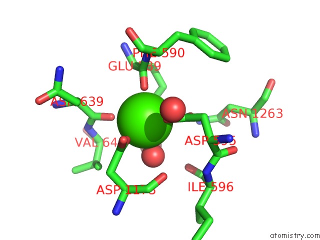
Mono view
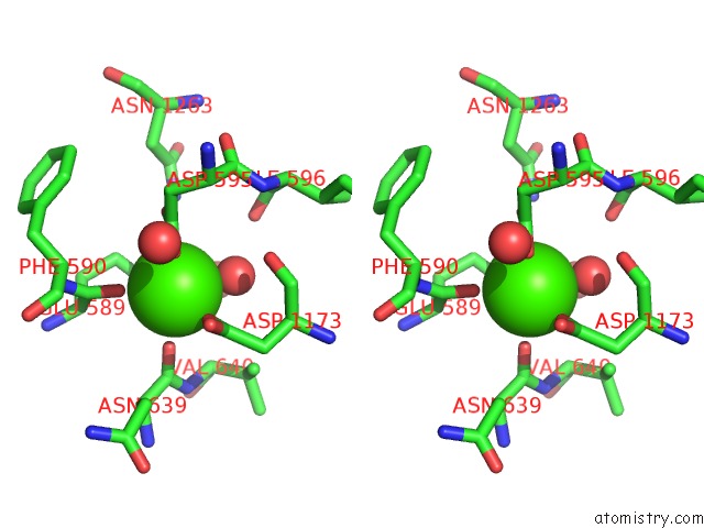
Stereo pair view
Mono view
Stereo pair view
A full contact list of Calcium with other atoms in the Ca binding
site number 1 of Crystal Structure of Truncated Alternansucrase From Leuconostoc Mesenteroides Nrrl B-1355 within 5.0Å range:
|
Calcium binding site 2 out of 2 in 6hvg
Go back to
Calcium binding site 2 out
of 2 in the Crystal Structure of Truncated Alternansucrase From Leuconostoc Mesenteroides Nrrl B-1355
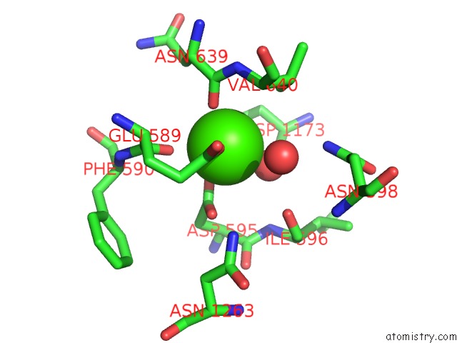
Mono view
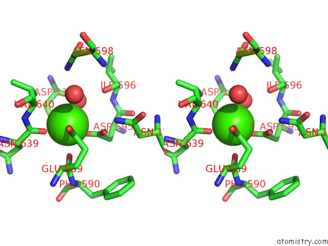
Stereo pair view
Mono view
Stereo pair view
A full contact list of Calcium with other atoms in the Ca binding
site number 2 of Crystal Structure of Truncated Alternansucrase From Leuconostoc Mesenteroides Nrrl B-1355 within 5.0Å range:
|
Reference:
M.Molina,
C.Moulis,
N.Monties,
S.Pizzut-Serin,
D.Guieysse,
S.Morel,
G.Cioci,
M.Remaud-Simeon.
Deciphering An Undecided Enzyme: Investigations of the Structural Determinants Involved in the Linkage Specificity of Alternansucrase Acs Catalysis 2019.
ISSN: ESSN 2155-5435
DOI: 10.1021/ACSCATAL.8B04510
Page generated: Wed Jul 9 14:43:01 2025
ISSN: ESSN 2155-5435
DOI: 10.1021/ACSCATAL.8B04510
Last articles
Ca in 7PKBCa in 7PI3
Ca in 7PJM
Ca in 7PHW
Ca in 7PIV
Ca in 7PI0
Ca in 7PIU
Ca in 7PC9
Ca in 7PH1
Ca in 7PEO