Calcium »
PDB 6q7o-6qps »
6q86 »
Calcium in PDB 6q86: Structure of Fucosylated D-Antimicrobial Peptide SB4 in Complex with the Fucose-Binding Lectin Pa-Iil at 2.008 Angstrom Resolution
Protein crystallography data
The structure of Structure of Fucosylated D-Antimicrobial Peptide SB4 in Complex with the Fucose-Binding Lectin Pa-Iil at 2.008 Angstrom Resolution, PDB code: 6q86
was solved by
S.Baeriswyl,
A.Stocker,
J.L.Reymond,
with X-Ray Crystallography technique. A brief refinement statistics is given in the table below:
| Resolution Low / High (Å) | 48.15 / 2.01 |
| Space group | P 31 2 1 |
| Cell size a, b, c (Å), α, β, γ (°) | 55.600, 55.600, 150.621, 90.00, 90.00, 120.00 |
| R / Rfree (%) | 15.7 / 19 |
Calcium Binding Sites:
The binding sites of Calcium atom in the Structure of Fucosylated D-Antimicrobial Peptide SB4 in Complex with the Fucose-Binding Lectin Pa-Iil at 2.008 Angstrom Resolution
(pdb code 6q86). This binding sites where shown within
5.0 Angstroms radius around Calcium atom.
In total 4 binding sites of Calcium where determined in the Structure of Fucosylated D-Antimicrobial Peptide SB4 in Complex with the Fucose-Binding Lectin Pa-Iil at 2.008 Angstrom Resolution, PDB code: 6q86:
Jump to Calcium binding site number: 1; 2; 3; 4;
In total 4 binding sites of Calcium where determined in the Structure of Fucosylated D-Antimicrobial Peptide SB4 in Complex with the Fucose-Binding Lectin Pa-Iil at 2.008 Angstrom Resolution, PDB code: 6q86:
Jump to Calcium binding site number: 1; 2; 3; 4;
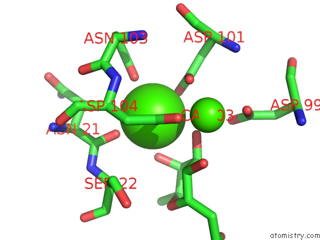
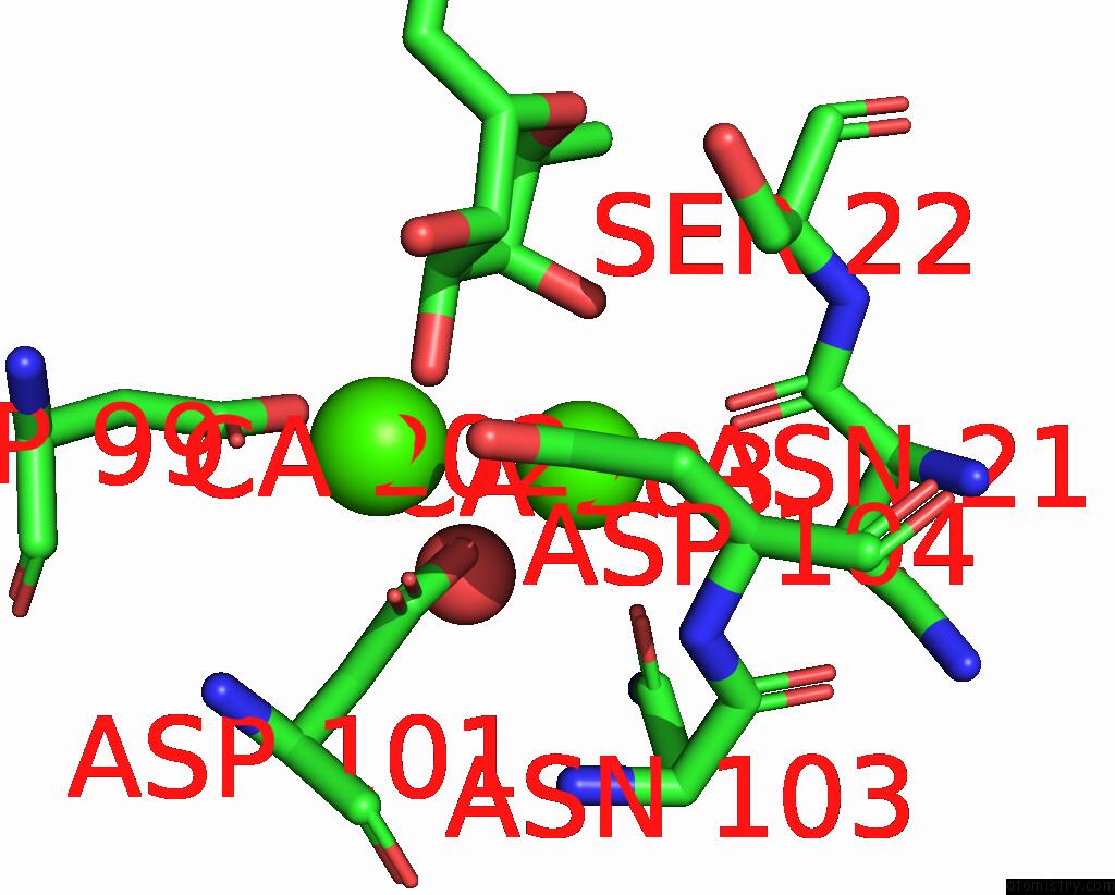
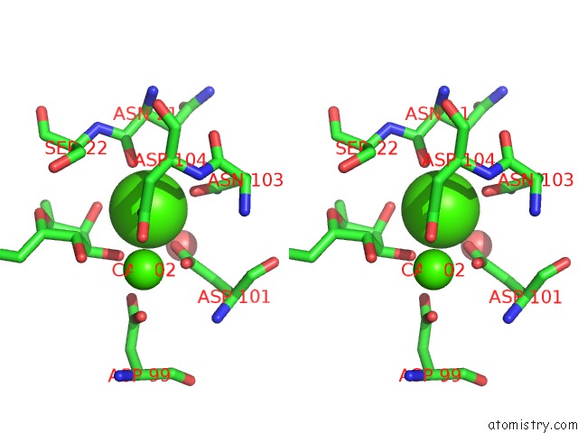
Calcium binding site 1 out of 4 in 6q86
Go back to
Calcium binding site 1 out
of 4 in the Structure of Fucosylated D-Antimicrobial Peptide SB4 in Complex with the Fucose-Binding Lectin Pa-Iil at 2.008 Angstrom Resolution
Mono view
Stereo pair view
Mono view
Stereo pair view
A full contact list of Calcium with other atoms in the Ca binding
site number 1 of Structure of Fucosylated D-Antimicrobial Peptide SB4 in Complex with the Fucose-Binding Lectin Pa-Iil at 2.008 Angstrom Resolution within 5.0Å range:
|
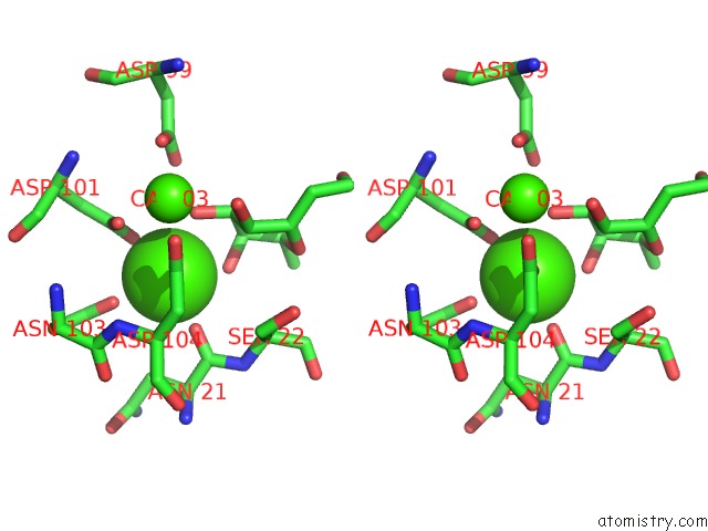
Calcium binding site 2 out of 4 in 6q86
Go back to
Calcium binding site 2 out
of 4 in the Structure of Fucosylated D-Antimicrobial Peptide SB4 in Complex with the Fucose-Binding Lectin Pa-Iil at 2.008 Angstrom Resolution

Mono view
Stereo pair view
Mono view
Stereo pair view
A full contact list of Calcium with other atoms in the Ca binding
site number 2 of Structure of Fucosylated D-Antimicrobial Peptide SB4 in Complex with the Fucose-Binding Lectin Pa-Iil at 2.008 Angstrom Resolution within 5.0Å range:
|
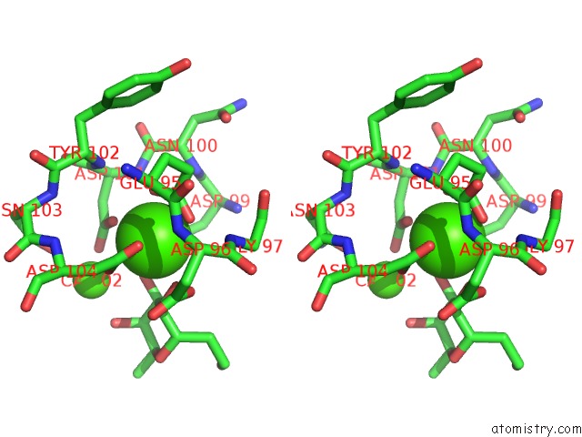
Calcium binding site 3 out of 4 in 6q86
Go back to
Calcium binding site 3 out
of 4 in the Structure of Fucosylated D-Antimicrobial Peptide SB4 in Complex with the Fucose-Binding Lectin Pa-Iil at 2.008 Angstrom Resolution
Mono view

Stereo pair view
Mono view
Stereo pair view
A full contact list of Calcium with other atoms in the Ca binding
site number 3 of Structure of Fucosylated D-Antimicrobial Peptide SB4 in Complex with the Fucose-Binding Lectin Pa-Iil at 2.008 Angstrom Resolution within 5.0Å range:
|
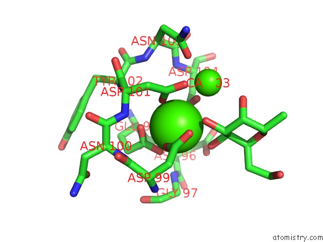
Calcium binding site 4 out of 4 in 6q86
Go back to
Calcium binding site 4 out
of 4 in the Structure of Fucosylated D-Antimicrobial Peptide SB4 in Complex with the Fucose-Binding Lectin Pa-Iil at 2.008 Angstrom Resolution
Mono view
Stereo pair view
Mono view
Stereo pair view
A full contact list of Calcium with other atoms in the Ca binding
site number 4 of Structure of Fucosylated D-Antimicrobial Peptide SB4 in Complex with the Fucose-Binding Lectin Pa-Iil at 2.008 Angstrom Resolution within 5.0Å range:
|
Reference:
S.Baeriswyl,
B.H.Gan,
T.N.Siriwardena,
R.Visini,
M.Robadey,
S.Javor,
A.Stocker,
T.Darbre,
J.L.Reymond.
X-Ray Crystal Structures of Short Antimicrobial Peptides As Pseudomonas Aeruginosa Lectin B Complexes. Acs Chem.Biol. V. 14 758 2019.
ISSN: ESSN 1554-8937
PubMed: 30830745
DOI: 10.1021/ACSCHEMBIO.9B00047
Page generated: Wed Jul 9 17:06:58 2025
ISSN: ESSN 1554-8937
PubMed: 30830745
DOI: 10.1021/ACSCHEMBIO.9B00047
Last articles
Cl in 5L2NCl in 5L1E
Cl in 5L2K
Cl in 5L0K
Cl in 5L2M
Cl in 5L2J
Cl in 5L0T
Cl in 5L0S
Cl in 5L0R
Cl in 5L0E